File:Frazer1914 fig02.jpg
From Embryology

Size of this preview: 559 × 600 pixels. Other resolution: 1,000 × 1,073 pixels.
Original file (1,000 × 1,073 pixels, file size: 261 KB, MIME type: image/jpeg)
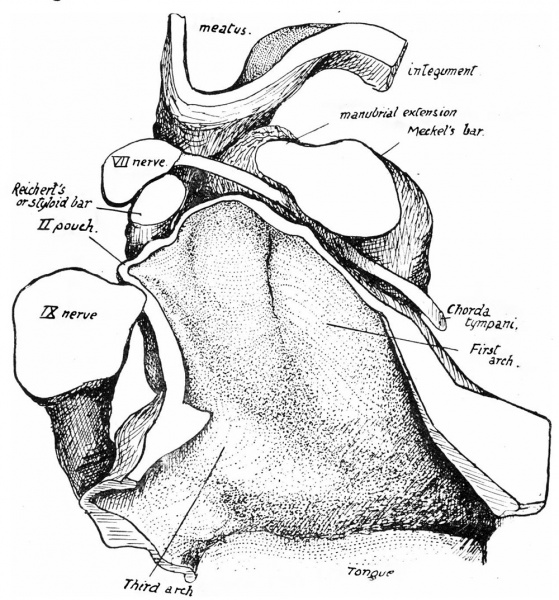
Fig. 2. Floor of left recess 16 mm Embryo
Floor of left recess, roof removed. From model (magn. 100 X 5/6). A portion of the margin of the tongue is seen below.
| Historic Disclaimer - information about historic embryology pages |
|---|
 |
Reference
Frazer JE. The second visceral arch and groove in the tubo-tympanic region. (1914) J Anat Physiol. 48(4): 391-408. PMID 17233005
Cite this page: Hill, M.A. (2024, May 20) Embryology Frazer1914 fig02.jpg. Retrieved from https://embryology.med.unsw.edu.au/embryology/index.php/File:Frazer1914_fig02.jpg
- © Dr Mark Hill 2024, UNSW Embryology ISBN: 978 0 7334 2609 4 - UNSW CRICOS Provider Code No. 00098G
File history
Click on a date/time to view the file as it appeared at that time.
| Date/Time | Thumbnail | Dimensions | User | Comment | |
|---|---|---|---|---|---|
| current | 06:00, 9 January 2017 | | 1,000 × 1,073 (261 KB) | Z8600021 (talk | contribs) | |
| 06:00, 9 January 2017 | 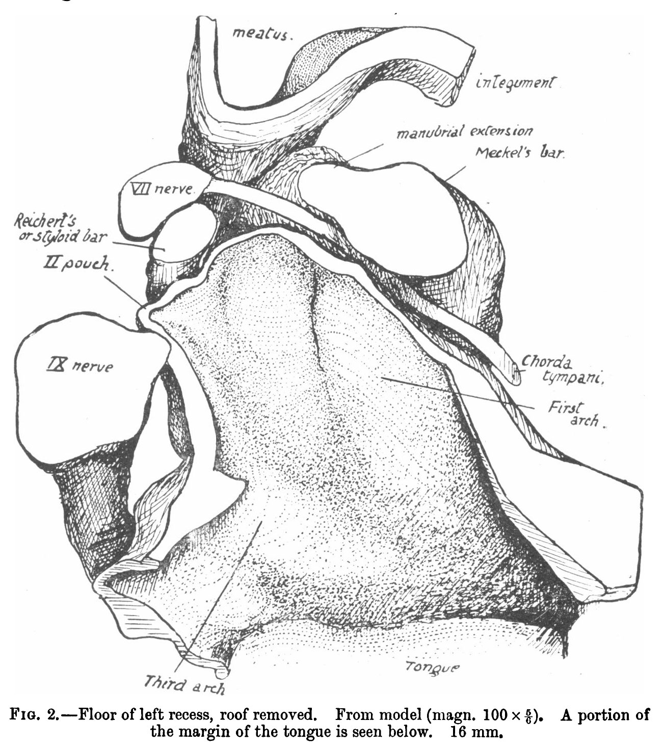 | 1,303 × 1,470 (352 KB) | Z8600021 (talk | contribs) |
You cannot overwrite this file.
File usage
The following 3 pages use this file: