File:Frazer1914 fig02.jpg

Original file (1,000 × 1,073 pixels, file size: 261 KB, MIME type: image/jpeg)
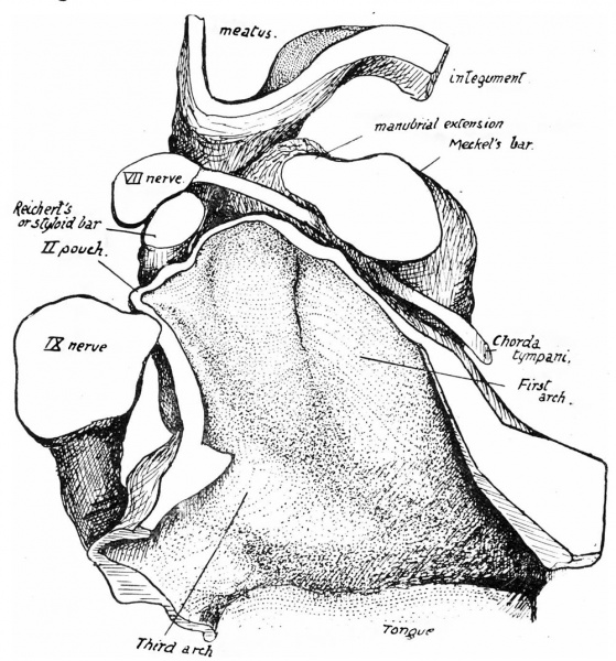
Fig. 2. Floor of left recess 16 mm Embryo
Floor of left recess, roof removed. From model (magn. 100 X 5/6). A portion of the margin of the tongue is seen below.
On removing the roof of the recess and thus exposing the floor, the disposition of the arches becomes apparent. fig. 2 is a drawing of the floor of the left recess in an embryo of 16 mm. exposed in this way, and is of interest because the key to subsequent changes is to be found in the conditions present at this stage.
The front part of the field is formed by structures of the first arch, among them Meckel’s bar passing downwards and inwards, and behind this a deep but narrow sulcus marks the situation of the first visceral groove ending externally in the first lateral pouch, just above which the chorda tympani runs forward. The second lateral pouch is apparent behind the styloid bar (Reichert’s bar), between this and the glossopharyngeal, which is closely applied to the wall of the recess here: this is the definite position of the second pouch, shown also in fig. 1 in the 12-mm. specimen, and is the position maintained by it until the growth of the cartilaginous auditory capsule effects a separation between it and the nerve.
| Historic Disclaimer - information about historic embryology pages |
|---|
 |
- Links: Fig 1 | Fig 2 | Fig 3 | Fig 4 | Fig 5 | Fig 6 | 1914 Frazer | Pharyngeal arches | Historic Embryology Papers
Reference
Frazer JE. The second visceral arch and groove in the tubo-tympanic region. (1914) J Anat Physiol. 48(4): 391-408. PMID 17233005
Cite this page: Hill, M.A. (2024, April 27) Embryology Frazer1914 fig02.jpg. Retrieved from https://embryology.med.unsw.edu.au/embryology/index.php/File:Frazer1914_fig02.jpg
- © Dr Mark Hill 2024, UNSW Embryology ISBN: 978 0 7334 2609 4 - UNSW CRICOS Provider Code No. 00098G
File history
Click on a date/time to view the file as it appeared at that time.
| Date/Time | Thumbnail | Dimensions | User | Comment | |
|---|---|---|---|---|---|
| current | 06:00, 9 January 2017 | | 1,000 × 1,073 (261 KB) | Z8600021 (talk | contribs) | |
| 06:00, 9 January 2017 | 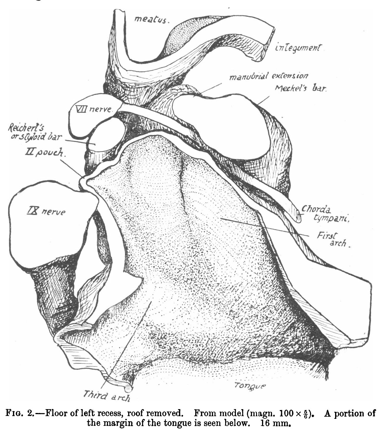 | 1,303 × 1,470 (352 KB) | Z8600021 (talk | contribs) |
You cannot overwrite this file.
File usage
The following 3 pages use this file: