File:Fetal 10wk urogenital 4.jpg: Difference between revisions
From Embryology
No edit summary |
No edit summary |
||
| Line 5: | Line 5: | ||
This stage of development is after the embryonic period (up to week 8) but still only 2 weeks into early fetal development. | This stage of development is after the embryonic period (up to week 8) but still only 2 weeks into early fetal development. | ||
Section | Section D is the most midline of all sections. Planes A, B, C and D move towards the midline. | ||
Original file name: | Original file name: H10wkUrogenAL.jpg http://embryology.med.unsw.edu.au/wwwhuman/Hum10wk/HumUrogen.htm | ||
{{Template:10wkFetus}} | |||
[[Category:Renal]] [[Category:Genital]] [[Category:Musculoskeletal]] | |||
Revision as of 15:56, 27 April 2010
Human Fetus
female, 10 week, 40 mm CRL, early fetal, sagittal section, pelvic region
This stage of development is after the embryonic period (up to week 8) but still only 2 weeks into early fetal development.
Section D is the most midline of all sections. Planes A, B, C and D move towards the midline.
Original file name: H10wkUrogenAL.jpg http://embryology.med.unsw.edu.au/wwwhuman/Hum10wk/HumUrogen.htm
Related Images
Fetus (week 10) Planes A (most lateral), B (lateral), C (medial) and D (midline) from lateral towards the midline.
- Human Fetus - most lateral | lateral | medial | midline
- Head - most lateral | lateral | medial | midline
- Cerebellum - most lateral | lateral | medial | midline
- Urogenital Unlabelled - most lateral | lateral | medial | midline
- Urogenital Labelled - most lateral | lateral | medial | midline
- Large Images - midline
- Image Source: UNSW Embryology, no reproduction without permission.
File history
Click on a date/time to view the file as it appeared at that time.
| Date/Time | Thumbnail | Dimensions | User | Comment | |
|---|---|---|---|---|---|
| current | 17:58, 28 May 2011 |  | 800 × 600 (105 KB) | S8600021 (talk | contribs) | |
| 17:55, 28 May 2011 | 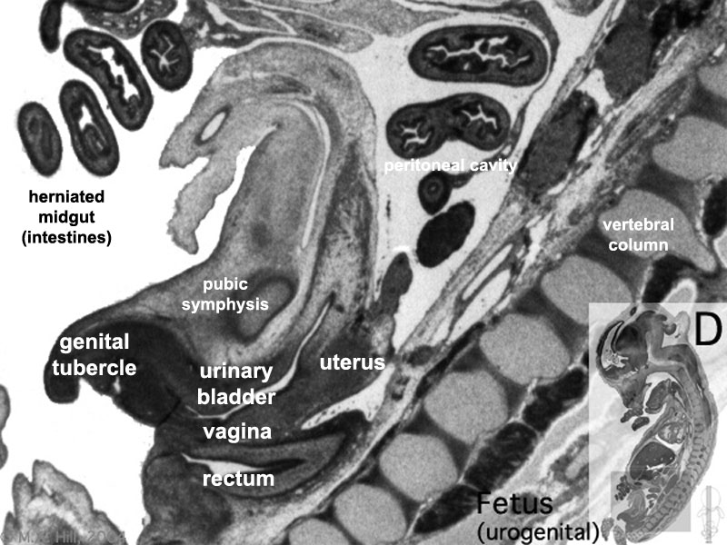 | 800 × 600 (105 KB) | S8600021 (talk | contribs) | relabeled and increased overall size of image. | |
| 22:58, 20 September 2009 | 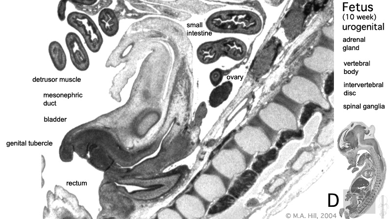 | 800 × 450 (125 KB) | S8600021 (talk | contribs) | These are images from an early fetus (female, 10 week, 40 mm). This stage of development is after the embryonic period (up to week 8) but still only 2 weeks into early fetal development. Section A is the most sagittal (lateral towards right) of all sectio |
You cannot overwrite this file.
File usage
The following 14 pages use this file:
- 2009 Lecture 15
- 2010 Lab 8
- 2010 Lecture 15
- 2011 Lab 8 - Fetal
- ANAT2241 Urinary System
- ANAT2341 Lab 8 - Fetal
- BGDB Sexual Differentiation - Fetal
- Fetal Development - 10 Weeks
- Genital - Female Development
- Lecture - Renal Development
- Renal System - Fetal
- Renal System Development
- Renal System Histology
- Urinary Bladder Development