File:Female genital and ureter abnormality 01.jpg

Original file (766 × 732 pixels, file size: 86 KB, MIME type: image/jpeg)
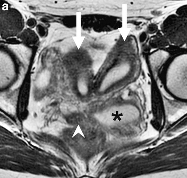
Female Genital and Ureter Abnormality
Uterine didelphys, obstructed hemivagina, and ectopic ureter on MR imaging in a 17-year-old girl.
a Axial T2-W image demonstrates two widely separate uterine horns (large arrows), an obstructed left hemivagina distended with fluid (asterisk), and a nondilated right hemivagina (arrowhead).
- Links: Axial T2-W image 1 | Axial T2-weighted image 2 | Coronal T2-W image | Genital System - Abnormalities | Renal System - Abnormalities
Reference
<pubmed>19924410</pubmed>| PMC2817805
Pediatr Radiol. 2010 March; 40(3): 358–360. Published online 2009 November 19. doi: 10.1007/s00247-009-1454-8.
Copyright
Copyright © The Author(s) 2009
Open Access This article is distributed under the terms of the Creative Commons Attribution Noncommercial License which permits any noncommercial use, distribution, and reproduction in any medium, provided the original author(s) and source are credited. Original file name: Fig. 1a 247_2009_1454_Fig1.jpg
Cite this page: Hill, M.A. (2024, April 27) Embryology Female genital and ureter abnormality 01.jpg. Retrieved from https://embryology.med.unsw.edu.au/embryology/index.php/File:Female_genital_and_ureter_abnormality_01.jpg
- © Dr Mark Hill 2024, UNSW Embryology ISBN: 978 0 7334 2609 4 - UNSW CRICOS Provider Code No. 00098G
File history
Click on a date/time to view the file as it appeared at that time.
| Date/Time | Thumbnail | Dimensions | User | Comment | |
|---|---|---|---|---|---|
| current | 15:31, 27 April 2011 | | 766 × 732 (86 KB) | S8600021 (talk | contribs) | ==Female Genital and Ureter Abnormality-- Uterine didelphys, obstructed hemivagina, and ectopic ureter on MR imaging in a 17-year-old girl. a Axial T2-W image demonstrates two widely separate uterine horns (large arrows), an obstructed left hemivagina di |
You cannot overwrite this file.
File usage
The following page uses this file: