File:Fawcett1913 fig04.jpg
Original file (928 × 664 pixels, file size: 122 KB, MIME type: image/jpeg)
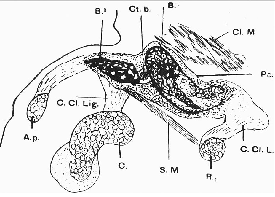
Fig. 4. Semi-schematic drawing of semi-coronal section of right clavicle of an 18 mm embryo
All the parts shown save the subclavius muscle were shown in the section drawn, but the subelavitus was added from its appearance in several sections. Notice how the sternal segment overrides the acromial segment ofthe clavicle.
A.p,acromion process; B.1,bone of sternal segment; B.2, bone of acromial segment; Ct.b.,pre- cartilaginous bridge unossified and connecting acromial and sternal segments; C. Cl. Lig., coraco-clavicular ligament; C, coracoid process; Cl.M., cleido-mastoid muscle; C.Cl.L.,costo-clavicular ligament; R.1, first rib; S. M., subclavius muscle.
Clavicle Links: Fig 1 | Fig 2 | Fig 3 | Fig 4 | Fig 5 | Fig 6 | Fig 7 | Fig 8 | 1913 Clavicle
- Edward Fawcett Links: 1906 Palate | 1910 Head | 1910 Sphenoid | 1911 Maxilla, vomer, and paraseptal cartilages | 1913 Clavicle | 1930 Mandible | Fawcett image | Edward Fawcett
Cite this page: Hill, M.A. (2024, April 28) Embryology Fawcett1913 fig04.jpg. Retrieved from https://embryology.med.unsw.edu.au/embryology/index.php/File:Fawcett1913_fig04.jpg
- © Dr Mark Hill 2024, UNSW Embryology ISBN: 978 0 7334 2609 4 - UNSW CRICOS Provider Code No. 00098G
File history
Click on a date/time to view the file as it appeared at that time.
| Date/Time | Thumbnail | Dimensions | User | Comment | |
|---|---|---|---|---|---|
| current | 14:40, 27 December 2014 | | 928 × 664 (122 KB) | Z8600021 (talk | contribs) | |
| 14:37, 27 December 2014 | 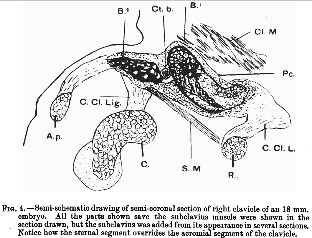 | 1,059 × 809 (177 KB) | Z8600021 (talk | contribs) | {{Fawcett1913 figures}} |
You cannot overwrite this file.
File usage
The following page uses this file: