File:Eisendrath1925 fig16.jpg: Difference between revisions
From Embryology
No edit summary |
mNo edit summary |
||
| (2 intermediate revisions by the same user not shown) | |||
| Line 1: | Line 1: | ||
==Fig. 16. Pyelographic findings in Case III== | |||
Note mesially directed calyces on both sides; also howfright pelvis extends across front of body of third lumbar vertebra. Note unusual form of both pelves. | |||
===Reference=== | |||
{{Ref-Eisendrath1925}} | |||
{{Footer}} | |||
[[Category:Historic Embryology]][[Category:Renal]][[Category:Abnormal Development]][[Category:1920's]] | |||
Latest revision as of 18:00, 7 September 2017
Fig. 16. Pyelographic findings in Case III
Note mesially directed calyces on both sides; also howfright pelvis extends across front of body of third lumbar vertebra. Note unusual form of both pelves.
Reference
Eisendrath DN Phifer FM and Culver HB. Horseshoe Kidney (1925) Ann Surg. 82(5): 735-64. PubMed 17865363
Cite this page: Hill, M.A. (2024, April 27) Embryology Eisendrath1925 fig16.jpg. Retrieved from https://embryology.med.unsw.edu.au/embryology/index.php/File:Eisendrath1925_fig16.jpg
- © Dr Mark Hill 2024, UNSW Embryology ISBN: 978 0 7334 2609 4 - UNSW CRICOS Provider Code No. 00098G
File history
Click on a date/time to view the file as it appeared at that time.
| Date/Time | Thumbnail | Dimensions | User | Comment | |
|---|---|---|---|---|---|
| current | 17:59, 7 September 2017 | 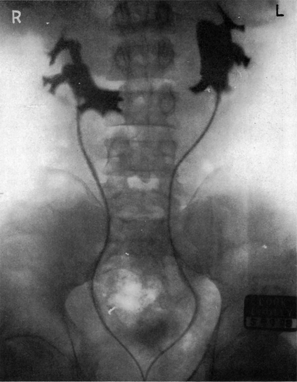 | 1,000 × 1,287 (76 KB) | Z8600021 (talk | contribs) | |
| 17:59, 7 September 2017 | 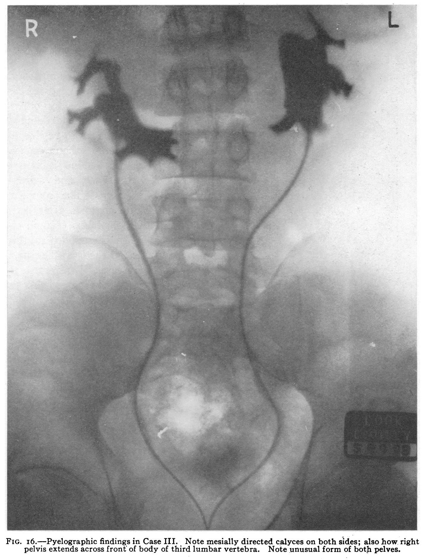 | 1,422 × 1,881 (322 KB) | Z8600021 (talk | contribs) |
You cannot overwrite this file.
File usage
The following 2 pages use this file: