File:Early Heart Tube (Lateral).jpg
From Embryology
.jpg&diff=prev&oldid=16958)
.jpg&direction=prev&oldid=16958)
.jpg)
.jpg&diff=cur&oldid=16958)
.jpg&direction=next&oldid=16958)
.jpg&diff=next&oldid=16958)

Size of this preview: 800 × 517 pixels. Other resolution: 1,504 × 972 pixels.
Original file (1,504 × 972 pixels, file size: 110 KB, MIME type: image/jpeg)
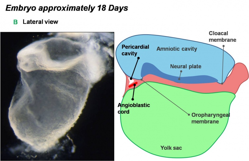
Image Source: Scanning electron micrographs of the Carnegie stages of the early human embryos are reproduced with the permission of Prof Kathy Sulik, from embryos collected by Dr. Vekemans and Tania Attié-Bitach. Images are for educational purposes only and cannot be reproduced electronically or in writing without permission.
Angiogenesis throughout the embryo allows for the development of angioblastic cords in the cardiogenic mesoderm of the embryo.
File history
Click on a date/time to view the file as it appeared at that time.
| Date/Time | Thumbnail | Dimensions | User | Comment | |
|---|---|---|---|---|---|
| current | 10:38, 14 March 2010 | | 1,504 × 972 (110 KB) | Z3212774 (talk | contribs) | category:Heart ILP Angiogenesis throughout the embryo allows for the development of angioblastic cords in the cardiogenic mesoderm of the embryo. |
You cannot overwrite this file.
.jpg&oldid=16958)