File:Duval1889 plate09.jpg
Original file (3,498 × 2,797 pixels, file size: 1.39 MB, MIME type: image/jpeg)
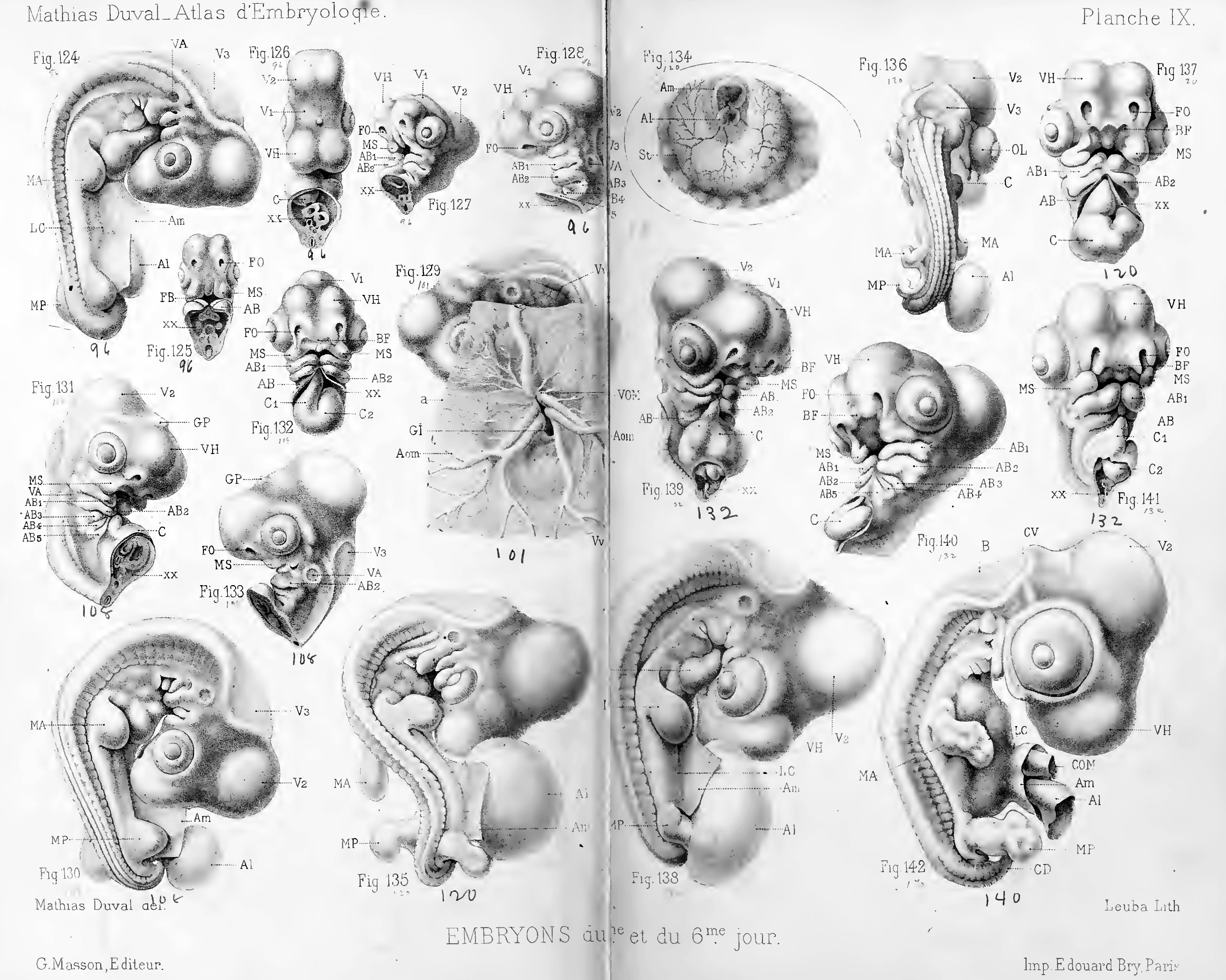
Plate 9 Chicken Embryo Day 5 to 6
This plate shows the chicken embryo.
This plate represents the embryo in the 5 and 6 incubation days (over 96 to 140 hour).
In previous boards, the embryo was represented seen by transparency; tnais from the end of the fourth day, the body of the embryo becomes too thick to make it possible, subsequently, to examine well by transparency, especially the outside then modeled the body draws very clearly the parties embryonic.
Therefore, in the stage IX and X, the embryos are represented by modeling, that is to say not seen by transparency (transmitted light), but the reflected light.
Reference
Duval M. Atlas d'embryologie (1889) G. Masson, Libraire De L'académie De Médecine Paris, France.
Cite this page: Hill, M.A. (2024, April 28) Embryology Duval1889 plate09.jpg. Retrieved from https://embryology.med.unsw.edu.au/embryology/index.php/File:Duval1889_plate09.jpg
- © Dr Mark Hill 2024, UNSW Embryology ISBN: 978 0 7334 2609 4 - UNSW CRICOS Provider Code No. 00098G
File history
Click on a date/time to view the file as it appeared at that time.
| Date/Time | Thumbnail | Dimensions | User | Comment | |
|---|---|---|---|---|---|
| current | 11:26, 19 June 2016 | | 3,498 × 2,797 (1.39 MB) | Z8600021 (talk | contribs) |
You cannot overwrite this file.
File usage
The following page uses this file: