File:Dextrocardia heart position.jpg: Difference between revisions
mNo edit summary |
mNo edit summary |
||
| Line 2: | Line 2: | ||
Anatomical position of the heart in the thorax. | Anatomical position of the heart in the thorax. | ||
During the inspection of the pulmonary trunk, a large ductus arteriosus (Botallo's ductus) was found. The heart apex was positioned along the heart axis and was turned to the right side, thus demonstrating the dextrocardia. | During the inspection of the pulmonary trunk, a large {{ductus arteriosus}} (Botallo's ductus) was found. The heart apex was positioned along the heart axis and was turned to the right side, thus demonstrating the dextrocardia. | ||
* '''1''' - pulmonary trunk | * '''1''' - pulmonary trunk | ||
| Line 13: | Line 13: | ||
* '''8''' - hepatic tissue covering the inferior vena cava (IVC) | * '''8''' - hepatic tissue covering the inferior vena cava (IVC) | ||
:'''Links:''' {{cardiac abnormalities}} | |||
===Reference=== | ===Reference=== | ||
| Line 21: | Line 20: | ||
====Copyright==== | ====Copyright==== | ||
All the content of the journal, except where otherwise noted, is licensed under a [http://creativecommons.org/licenses/by-nc/3.0/ Creative Commons License]. | All the content of the journal, except where otherwise noted, is licensed under a [http://creativecommons.org/licenses/by-nc/3.0/ Creative Commons License]. | ||
Original File Name: En_a13fig01.jpg female child, probably one year of age, which belonged to the Laboratory of Anatomy of the Campus of São José dos Campos - UNESP. | |||
{{Footer}} | {{Footer}} | ||
[[Category:Cardiovascular]] [[Category:Heart]] [[Category:Abnormal Development]] | [[Category:Cardiovascular]] [[Category:Heart]] [[Category:Abnormal Development]] | ||
Revision as of 10:19, 2 August 2019
Dextrocardia
Anatomical position of the heart in the thorax.
During the inspection of the pulmonary trunk, a large ductus arteriosus (Botallo's ductus) was found. The heart apex was positioned along the heart axis and was turned to the right side, thus demonstrating the dextrocardia.
- 1 - pulmonary trunk
- 2 - ascending aorta
- 3 - superior vena cava (SVC)
- 4 - brachiocephalic trunk
- 5 - left common carotid artery
- 6 - left subclavial artery
- 7 - thoracic aorta
- 8 - hepatic tissue covering the inferior vena cava (IVC)
Reference
Faig-Leite FS & Faig-Leite H. (2008). Anatomy of a dextrocardia case with situs solitus. Arq. Bras. Cardiol. , 91, e64-6. PMID: 19142355
Copyright
All the content of the journal, except where otherwise noted, is licensed under a Creative Commons License.
Original File Name: En_a13fig01.jpg female child, probably one year of age, which belonged to the Laboratory of Anatomy of the Campus of São José dos Campos - UNESP.
Cite this page: Hill, M.A. (2024, May 26) Embryology Dextrocardia heart position.jpg. Retrieved from https://embryology.med.unsw.edu.au/embryology/index.php/File:Dextrocardia_heart_position.jpg
- © Dr Mark Hill 2024, UNSW Embryology ISBN: 978 0 7334 2609 4 - UNSW CRICOS Provider Code No. 00098G
File history
Click on a date/time to view the file as it appeared at that time.
| Date/Time | Thumbnail | Dimensions | User | Comment | |
|---|---|---|---|---|---|
| current | 00:48, 7 August 2010 | 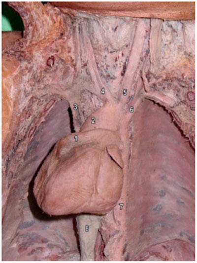 | 400 × 533 (49 KB) | S8600021 (talk | contribs) | ==Dextrocardia== Anatomical position of the heart in the thorax. During the inspection of the pulmonary trunk, a large ductus arteriosus (Botallo's ductus) was found. The heart apex was positioned along the heart axis and was turned to the right side, th |
You cannot overwrite this file.
File usage
The following 4 pages use this file: