File:Congdon1922-17.jpg
From Embryology

Size of this preview: 800 × 329 pixels. Other resolution: 1,000 × 411 pixels.
Original file (1,000 × 411 pixels, file size: 55 KB, MIME type: image/jpeg)
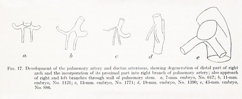
Fig. 17. Development of the pulmonary artery and ductus arteriosus
Showing degeneration of distal part of right arch and the incorporation of its proximal part into right branch of pulmonary artery; also approach of right and left branches through wall of pulmonary stem
a, 7-mm. embryo, No. 617
b, 11-mm. embryo, No. 1121
c, 13-mm. embryo, No. 1771
d, 18-mm. embryo, No. 1390
e, 43-mm. embryo, No. 886
| Historic Disclaimer - information about historic embryology pages |
|---|
 |
File history
Click on a date/time to view the file as it appeared at that time.
| Date/Time | Thumbnail | Dimensions | User | Comment | |
|---|---|---|---|---|---|
| current | 15:30, 1 September 2012 | 1,000 × 411 (55 KB) | Z8600021 (talk | contribs) | ||
| 17:38, 7 May 2011 | 1,144 × 428 (64 KB) | S8600021 (talk | contribs) | ==Fig. 17. Development of the pulmonary artery and ductus arteriosus== Showing degeneration of distal part of right arch and the incorporation of its proximal part into right branch of pulmonary artery; also approach of right and left branches through wa |
You cannot overwrite this file.
File usage
The following page uses this file: