File:Congdon1922-1-16.jpg
From Embryology

Size of this preview: 588 × 600 pixels. Other resolution: 980 × 1,000 pixels.
Original file (980 × 1,000 pixels, file size: 157 KB, MIME type: image/jpeg)
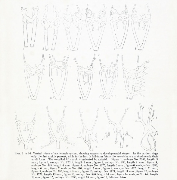
Figs. 1 to 16. Ventral views of aortic-arch system
Showing successive developmental stages. In the earliest stage only the first arch is present, while in the last (a full-term fetus) the vessels have acquired nearly their adult form. The so-called fifth arch is indicated by asterisk.
- Figure 1, embryo No. 2053, length 3 mm.
- Figure 2, embryo No. 12016, length 3 mm.
- Figure 3, embryo No. 836, length 4 mm.
- Figure 4, embryo No. 588, length 4 mm.
- Figure 5, embryo No. 1075, length 6 mm.
- Figure 6, embryo No. 1380, length 6 mm.
- Figure 7, embryo No. 810, length 5 mm.
- Figure 8, embryo No. 617, length 7 mm.
- Figure 9, embryo No. 792, length 8 mm.
- Figure 10, embryo No. 1121, length 11 mm.
- Figure 12, embryo No. 1771, length 13 mm.
- Figure 13, embryo No. 940, length 14 mm.
- Figure 14, embryo No. 74, length 16 mm.
- Figure 15, embryo No. 1390, length 18 mm.
- Figure 16, full-term fetus.
| Historic Disclaimer - information about historic embryology pages |
|---|
| Pages where the terms "Historic" (textbooks, papers, people, recommendations) appear on this site, and sections within pages where this disclaimer appears, indicate that the content and scientific understanding are specific to the time of publication. This means that while some scientific descriptions are still accurate, the terminology and interpretation of the developmental mechanisms reflect the understanding at the time of original publication and those of the preceding periods, these terms, interpretations and recommendations may not reflect our current scientific understanding. (More? Embryology History | Historic Embryology Papers) |
File history
Click on a date/time to view the file as it appeared at that time.
| Date/Time | Thumbnail | Dimensions | User | Comment | |
|---|---|---|---|---|---|
| current | 08:22, 1 September 2012 |  | 980 × 1,000 (157 KB) | Z8600021 (talk | contribs) | |
| 17:49, 7 May 2011 |  | 888 × 891 (74 KB) | S8600021 (talk | contribs) | ==Figs. 1 to 16. Ventral views of aortic-arch system== Showing successive developmental stages. In the earliest stage only the first arch is present, while in the last (a full-term fetus) the vessels have acquired nearly their adult form. The so-called f |
You cannot overwrite this file.
File usage
The following page uses this file:
