File:Comparative brain anatomy frog-dog.jpg: Difference between revisions
From Embryology
No edit summary |
|||
| (3 intermediate revisions by the same user not shown) | |||
| Line 3: | Line 3: | ||
These 2 models are viewed from a similar direction and components have similar colour codings (the comparative sizes are not to scale). | These 2 models are viewed from a similar direction and components have similar colour codings (the comparative sizes are not to scale). | ||
Note the dog brain has, compared to other brain components, a larger telencephalon (cortex) and cerebellum than the frog brain. | Note: | ||
# Both brains have a large region devoted to smell (olfactory bulb). | |||
# Mammalian (dog) telencephalon (cortex) has grown over the the midbrain, visible in the amphibian (frog). | |||
# The dog brain (mammalian) has, compared to other brain components, a larger telencephalon (cortex) and cerebellum than the frog brain (amphibian). | |||
Latest revision as of 11:41, 15 February 2013
Comparative Brain Anatomy Frog and Dog
These 2 models are viewed from a similar direction and components have similar colour codings (the comparative sizes are not to scale).
Note:
- Both brains have a large region devoted to smell (olfactory bulb).
- Mammalian (dog) telencephalon (cortex) has grown over the the midbrain, visible in the amphibian (frog).
- The dog brain (mammalian) has, compared to other brain components, a larger telencephalon (cortex) and cerebellum than the frog brain (amphibian).
File history
Click on a date/time to view the file as it appeared at that time.
| Date/Time | Thumbnail | Dimensions | User | Comment | |
|---|---|---|---|---|---|
| current | 16:21, 13 March 2012 | 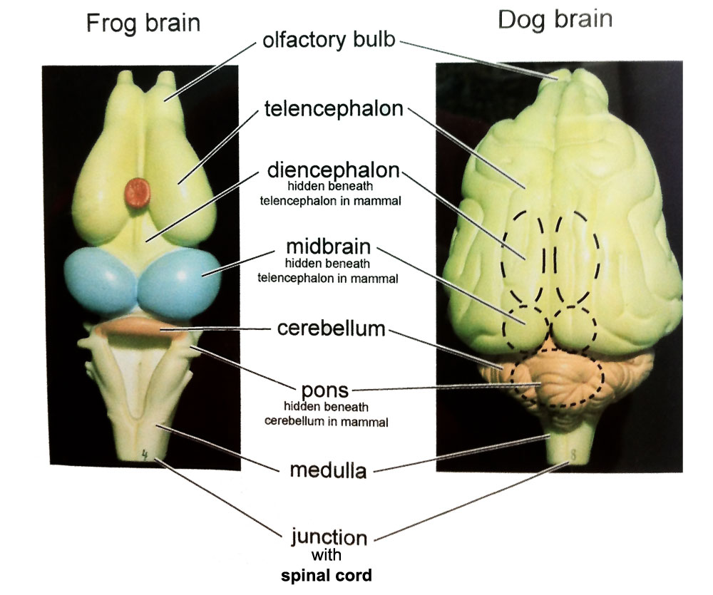 | 1,000 × 835 (112 KB) | Z8600021 (talk | contribs) | ==Comparative Brain Anatomy Frog and Dog== Category:Neural Category:Frog Category:Dog |
You cannot overwrite this file.
File usage
The following 4 pages use this file: