File:Cervical intersomitic vessels.png: Difference between revisions
From Embryology
mNo edit summary |
|||
| Line 1: | Line 1: | ||
==Mouse - Cervical Intersomitic Vessel== | ==Mouse - Cervical Intersomitic Vessel== | ||
'''A''' The various stages of cervical intersomitic vessel development can be segmented as visualized as surface renderings in the 16 somite mouse embryo ([[Mouse_Stages#Theiler_Stage_13|Theiler Stage 13]] - [[Mouse_Stages#Theiler_Stage_14|Theiler Stage 14]]) | * '''A''' The various stages of cervical intersomitic vessel development can be segmented as visualized as surface renderings in the 16 somite mouse embryo ([[Mouse_Stages#Theiler_Stage_13|Theiler Stage 13]] - [[Mouse_Stages#Theiler_Stage_14|Theiler Stage 14]]) The vessels along the right side of the embryo and surrounding somites 1 through 16 are labelled as: DA (red), ISA (pink), ISV (blue), VTA (vertebral artery), DLAV (dorsal longitudinal anastomotical vessel) and PNVP (green) (perineual vascular plexus) , ACV (anterior cardinal vein) and CCV (cyan) (common cardinal vein) , UV (dark pink), UV plexus (purple), and PCV (blue). Somites 1, 5, 10 and 15 are numbered as S-1, S-5, S-10 and S-15. | ||
* '''B''' Branches of PECAM-1 expression originating from the tips of the ISAs (yellow arrowheads) were observed to turn towards the location of the future PCV (posterior cardinal vein). | |||
The vessels along the right side of the embryo and surrounding somites 1 through 16 are labelled as: DA (red), ISA (pink), ISV (blue), VTA (vertebral artery), DLAV (dorsal longitudinal anastomotical vessel) and PNVP (green) (perineual vascular plexus) , ACV (anterior cardinal vein) and CCV (cyan) (common cardinal vein) , UV (dark pink), UV plexus (purple), and PCV (blue). Somites 1, 5, 10 and 15 are numbered as S-1, S-5, S-10 and S-15. | * '''C''' A second branch from the ISA was also observed to extend in a predominantly anterior direction (pink arrowheads) to connect up with other ISAs, eventually forming the DLAV. PECAM-1 expression along the location of the expected PCV was observed to lag development of the ISAs and is discontinuous (yellow arrowheads). | ||
* '''D''' The PNVP develops through remodelling of the VTA and DLAV. Branches initiate medially from the DLAV (pink arrowhead), begin to remodel into simple mesh (blue arrowhead), and eventually remodel into a fine structured capillary plexus surrounding the neural tube. Note at this stage that the first ISA has regressed. | |||
'''B''' Branches of PECAM-1 expression originating from the tips of the ISAs (yellow arrowheads) were observed to turn towards the location of the future PCV (posterior cardinal vein). | |||
'''C''' A second branch from the ISA was also observed to extend in a predominantly anterior direction (pink arrowheads) to connect up with other ISAs, eventually forming the DLAV. PECAM-1 expression along the location of the expected PCV was observed to lag development of the ISAs and is discontinuous (yellow arrowheads). | |||
'''D''' The PNVP develops through remodelling of the VTA and DLAV. Branches initiate medially from the DLAV (pink arrowhead), begin to remodel into simple mesh (blue arrowhead), and eventually remodel into a fine structured capillary plexus surrounding the neural tube. Note at this stage that the first ISA has regressed. | |||
Scale bars represent 100 microns. | Scale bars represent 100 microns. | ||
{| | |||
! Legend | |||
'''ACV''' - anterior cardinal vein | |- | ||
| | |||
'''CCV''' - common cardinal vein | * '''ACV''' - anterior cardinal vein | ||
* '''CCV''' - common cardinal vein | |||
'''DA''' - dorsal aorta | * '''DA''' - dorsal aorta | ||
* '''DLAV''' - dorsal longitudinal anastomotical vessel | |||
| | |||
* '''ISA''' - intersomitic artery | |||
* '''ISV''' - intersomitic vein | |||
* '''OA''' - omphalomesenteric artery | |||
* '''OV''' - omphalomesenteric vein | |||
| | |||
* '''PCV''' - posterior cardinal vein | |||
* '''PNVP''' - perineual vascular plexus | |||
* '''UA''' - umbilical artery | |||
* '''UV''' - umbilical vein | |||
|} | |||
{{Heart links}} | |||
===Reference=== | |||
<pubmed>18682734</pubmed>| [http://www.plosone.org/article/info:doi/10.1371/journal.pone.0002853 PLoS ONE] | <pubmed>18682734</pubmed>| [http://www.plosone.org/article/info:doi/10.1371/journal.pone.0002853 PLoS ONE] | ||
Copyright | ====Copyright==== | ||
© 2008 Walls et al. This is an open-access article distributed under the terms of the Creative Commons Attribution License, which permits unrestricted use, distribution, and reproduction in any medium, provided the original author and source are credited. | |||
Original File: http://www.plosone.org/article/slideshow.action?uri=info:doi/10.1371/journal.pone.0002853&imageURI=info:doi/10.1371/journal.pone.0002853.g009 | |||
[[Category:Heart]] [[Category:Cardiovascular]] [[Category:Mouse]] [[Category:Mouse E8.5]] [[Category:Mouse E9.0]] | [[Category:Heart]] [[Category:Cardiovascular]] [[Category:Mouse]] [[Category:Mouse E8.5]] [[Category:Mouse E9.0]] | ||
Revision as of 17:21, 22 August 2014
Mouse - Cervical Intersomitic Vessel
- A The various stages of cervical intersomitic vessel development can be segmented as visualized as surface renderings in the 16 somite mouse embryo (Theiler Stage 13 - Theiler Stage 14) The vessels along the right side of the embryo and surrounding somites 1 through 16 are labelled as: DA (red), ISA (pink), ISV (blue), VTA (vertebral artery), DLAV (dorsal longitudinal anastomotical vessel) and PNVP (green) (perineual vascular plexus) , ACV (anterior cardinal vein) and CCV (cyan) (common cardinal vein) , UV (dark pink), UV plexus (purple), and PCV (blue). Somites 1, 5, 10 and 15 are numbered as S-1, S-5, S-10 and S-15.
- B Branches of PECAM-1 expression originating from the tips of the ISAs (yellow arrowheads) were observed to turn towards the location of the future PCV (posterior cardinal vein).
- C A second branch from the ISA was also observed to extend in a predominantly anterior direction (pink arrowheads) to connect up with other ISAs, eventually forming the DLAV. PECAM-1 expression along the location of the expected PCV was observed to lag development of the ISAs and is discontinuous (yellow arrowheads).
- D The PNVP develops through remodelling of the VTA and DLAV. Branches initiate medially from the DLAV (pink arrowhead), begin to remodel into simple mesh (blue arrowhead), and eventually remodel into a fine structured capillary plexus surrounding the neural tube. Note at this stage that the first ISA has regressed.
Scale bars represent 100 microns.
| Legend | ||
|---|---|---|
|
|
|
Reference
<pubmed>18682734</pubmed>| PLoS ONE
Copyright
© 2008 Walls et al. This is an open-access article distributed under the terms of the Creative Commons Attribution License, which permits unrestricted use, distribution, and reproduction in any medium, provided the original author and source are credited.
File history
Click on a date/time to view the file as it appeared at that time.
| Date/Time | Thumbnail | Dimensions | User | Comment | |
|---|---|---|---|---|---|
| current | 11:56, 15 August 2009 | 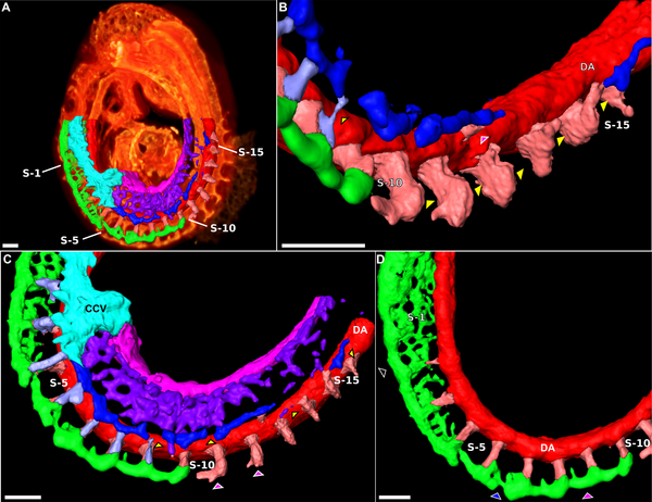 | 600 × 462 (308 KB) | S8600021 (talk | contribs) | (A) The various stages of cervical intersomitic vessel development can be segmented as visualized as surface renderings in the 16 somite mouse embryo. The vessels along the right side of the embryo and surrounding somites 1 through 16 are labelled as: D |
You cannot overwrite this file.
File usage
The following 4 pages use this file: