File:Carnegie stage 22 - pectoralis major muscle.jpg
Original file (1,965 × 1,019 pixels, file size: 313 KB, MIME type: image/jpeg)
Pectoralis Major Muscle
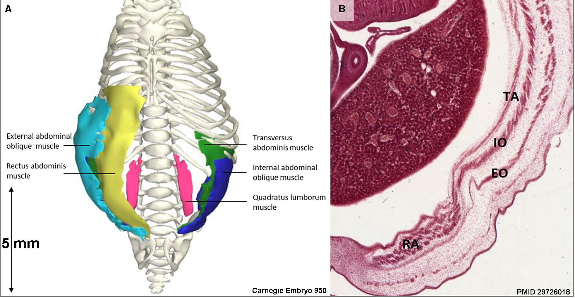
(A) Frontal view of the reconstructed pectoralis major portions of a stage 23 human embryo (56–60 days of development), specimen number 950. All other muscles are hidden. Depicted are three heads of the pectoralis major: clavicular head (light blue), sternocostal head (red) and abdominal head (yellow) and bones (white).
(B) Transverse section of the left shoulder region. The pectoralis major muscle is divided into three heads at this stage and is therefore even more partitioned than in adults (Gilroy et al. 2009). The tree heads of the left pectoralis muscle and the second rib and humerus of the same specimen as in (A) are tagged.
Legend: AH, abdominal head; CH, clavicular head; PM, pectoralis major muscle; SCH, sternocostal head. Scale bar: ~5 mm.
Reference
Warmbrunn MV, de Bakker BS, Hagoort J, Alefs-de Bakker PB & Oostra RJ. (2018). Hitherto unknown detailed muscle anatomy in an 8-week-old embryo. J. Anat. , , . PMID: 29726018 DOI.
Gilroy AM, MacPherson BR, Ross LM, et al. (2009) Atlas of Anatomy. Stuttgart: Thieme Medical Publishers.
Copyright
© 2018 The Authors. Journal of Anatomy published by John Wiley & Sons Ltd on behalf of Anatomical Society This is an open access article under the terms of the Creative Commons Attribution NonCommercial License, which permits use, distribution and reproduction in any medium, provided the original work is properly cited and is not used for commercial purposes.
Fig. 6 cropped relabelled with PMID, Embryo number and scale bar measurement.
Cite this page: Hill, M.A. (2024, April 27) Embryology Carnegie stage 22 - pectoralis major muscle.jpg. Retrieved from https://embryology.med.unsw.edu.au/embryology/index.php/File:Carnegie_stage_22_-_pectoralis_major_muscle.jpg
- © Dr Mark Hill 2024, UNSW Embryology ISBN: 978 0 7334 2609 4 - UNSW CRICOS Provider Code No. 00098G
File history
Click on a date/time to view the file as it appeared at that time.
| Date/Time | Thumbnail | Dimensions | User | Comment | |
|---|---|---|---|---|---|
| current | 11:43, 27 May 2018 | | 1,965 × 1,019 (313 KB) | Z8600021 (talk | contribs) | |
| 11:40, 27 May 2018 |  | 1,986 × 1,379 (460 KB) | Z8600021 (talk | contribs) | ==Pectoralis major muscle== (A) Frontal view of the reconstructed pectoralis major portions of a stage 23 human embryo (56–60 days of development), specimen number 950. All other muscles are hidden. Depicted are three heads of the pectoralis major:... |
You cannot overwrite this file.
File usage
The following page uses this file: