File:Bryce1908 fig05.jpg: Difference between revisions
(==Figure 5. == {{Historic Disclaimer}} Category:Human) |
m (→Figure 5.) |
||
| Line 1: | Line 1: | ||
==Figure 5. == | ==Figure 5. Diagram to illustrate the condition of Entypy of the the Germinal Area== | ||
The trophoblast is represented by continuous black lines or masses, there entoderm by interrupted lines, the embryonic ectoderm, and in certain figures the amniotic ectoderm, by epithelial cells. | |||
Each figure represents the blastodemic vesicle, a, of the rabbit ; b, of the mole ; c, of the bat ; d, of the mouse or rat ; e, of the guinea-pig ; f, of the kalong (I'teropus edulis). | |||
Ill the rnbbit (a) the cells of the embryonic knob early become arranged as an oiiithulial plate at the upjier pole of the blastocyst : the covering layer of trophoblaat (Riiiiuer's layer) disappears and it is exposed on the surface— there is do umiiiO'i'iiibryonic cavity. | |||
in Ihe mole (6) the embryonic plate is for a short time ioturned. The hallow is HUihI with trnpholilast cells which disappear, and the plate straightening out is uxiKmud uD the surface as in the rabbit. | |||
In the bat (ir) a more distinct cavity appears in the heart of the embryonic kaah. 'I'hu tliHir of this forma the embryonic ectoilerm which is necessarily at IJrst coni«vo (f'.f. Intumeil) ; the roof pereists as the primilive amnion. | |||
Ih the iiiuuao or rat [d) the covering laj er of tmphoblast roo5ng in the primitive niiiiilotii! unvity Iwconies greatly thickened, and ihe slightly intumed embi^onie plate (a jiuBhiMl Inwards as the blastocyst elongates into a tubular shape, until the layiTH (i-i'tiHlerm and ontD<lemi) arc apparently reversed in position. | |||
In till' )("lnca-)iig (i>) the same inpushing occurs, but the amnio-embryonic rudiment and the trophoblastic thJckciimg arc separated ; the amnio-embryonic vesicle remaiiiH a olosiil vesicle iiud forms the definitirr amniotic cavity. | |||
In I'tcrripUH (/) the conditions are mucli the same as in Cavia, bat the blaato | |||
uvNt I'l tins nmndcd and there is no greater intuming of the layers than occurs in | |||
llic i-arly phiwos of the mole. | |||
Revision as of 00:56, 3 November 2013
Figure 5. Diagram to illustrate the condition of Entypy of the the Germinal Area
The trophoblast is represented by continuous black lines or masses, there entoderm by interrupted lines, the embryonic ectoderm, and in certain figures the amniotic ectoderm, by epithelial cells.
Each figure represents the blastodemic vesicle, a, of the rabbit ; b, of the mole ; c, of the bat ; d, of the mouse or rat ; e, of the guinea-pig ; f, of the kalong (I'teropus edulis).
Ill the rnbbit (a) the cells of the embryonic knob early become arranged as an oiiithulial plate at the upjier pole of the blastocyst : the covering layer of trophoblaat (Riiiiuer's layer) disappears and it is exposed on the surface— there is do umiiiO'i'iiibryonic cavity.
in Ihe mole (6) the embryonic plate is for a short time ioturned. The hallow is HUihI with trnpholilast cells which disappear, and the plate straightening out is uxiKmud uD the surface as in the rabbit.
In the bat (ir) a more distinct cavity appears in the heart of the embryonic kaah. 'I'hu tliHir of this forma the embryonic ectoilerm which is necessarily at IJrst coni«vo (f'.f. Intumeil) ; the roof pereists as the primilive amnion.
Ih the iiiuuao or rat [d) the covering laj er of tmphoblast roo5ng in the primitive niiiiilotii! unvity Iwconies greatly thickened, and ihe slightly intumed embi^onie plate (a jiuBhiMl Inwards as the blastocyst elongates into a tubular shape, until the layiTH (i-i'tiHlerm and ontD<lemi) arc apparently reversed in position.
In till' )("lnca-)iig (i>) the same inpushing occurs, but the amnio-embryonic rudiment and the trophoblastic thJckciimg arc separated ; the amnio-embryonic vesicle remaiiiH a olosiil vesicle iiud forms the definitirr amniotic cavity.
In I'tcrripUH (/) the conditions are mucli the same as in Cavia, bat the blaato
uvNt I'l tins nmndcd and there is no greater intuming of the layers than occurs in
llic i-arly phiwos of the mole.
| Historic Disclaimer - information about historic embryology pages |
|---|
 |
File history
Click on a date/time to view the file as it appeared at that time.
| Date/Time | Thumbnail | Dimensions | User | Comment | |
|---|---|---|---|---|---|
| current | 00:49, 3 November 2013 | 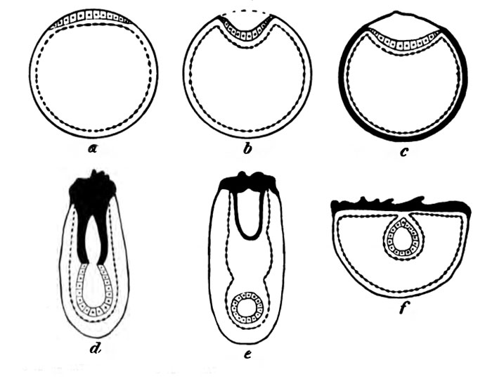 | 700 × 536 (0 bytes) | Z8600021 (talk | contribs) | ==Figure 5. == {{Historic Disclaimer}} Category:Human |
You cannot overwrite this file.
File usage
The following 2 pages use this file: