File:Braune 1877 plate 2 fig4.jpg
Original file (809 × 1,000 pixels, file size: 213 KB, MIME type: image/jpeg)
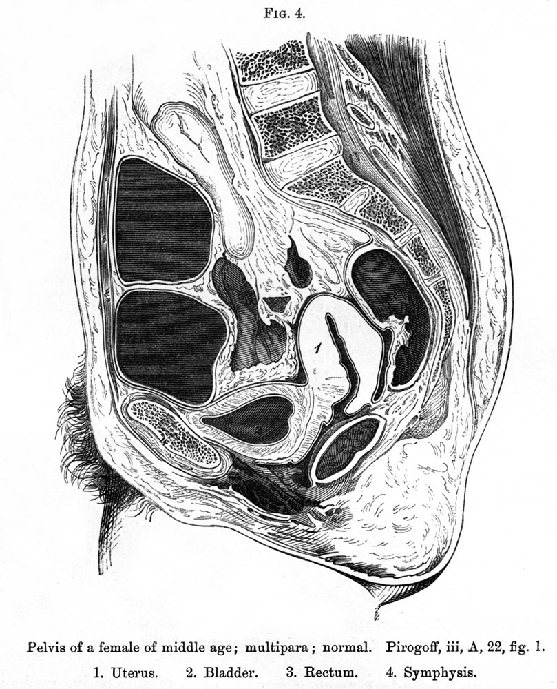
Plate 2 Sagittal Female Fig. 4. Pelvis of a female of middle age; multipara ; normal
Pirogoff, iii, A, 22, fig. 1. 1. Uterus. 2. Bladder. 3. Rectum. 4. Symphysis.
The uterus in fig. 4 with all its connections, was normal, and lay between the moderately distended bladder and rectum ; nor do coils of intestine lie behind the uterus in this section.
It will be noticed, therefore, that in the different degrees of distension of the bladder and rectum the uterus is always in the middle line between these viscera, whilst its position varies with its volume.
The uterus in this figure lies considerably deeper than in the foregoing ones. The conjugate diameter is 4.2 inches.
- Sagittal Female: Plate 2A | Plate 2B | Plate 2 Fig.1 | Plate 2 Fig.2 | Plate 2 Fig.3 | Plate 2 Fig.4 | Plate 2 Fig.5 | Plate 2 Fig.6 | Plate 2 Fig.7 | Topographical Anatomy (1877)
| Historic Disclaimer - information about historic embryology pages |
|---|
 |
- Braune Plates (1877): 1. Male - Sagittal body | 2. Female - Sagittal body | 3. Obliquely transverse head | 4. Transverse internal ear | 5. Transverse head | 6. Transverse neck | 7. Transverse neck and shoulders | 8. Transverse level first dorsal vertebra | 9. Transverse thorax level of third dorsal vertebra | 10. Transverse level aortic arch and fourth dorsal vertebra | 11. Transverse level of the bulbus aortae and sixth dorsal vertebra | 12. Transverse level of mitral valve and eighth dorsal vertebra | 13. Transverse level of heart apex and ninth dorsal vertebra | 14. Transverse liver stomach spleen at level of eleventh dorsal vertebra | 15. Transverse pancreas and kidneys at level of L1 vertebra | 16. Transverse through transverse colon at level of intervertebral space between L3 L4 vertebra | 17. Transverse pelvis at level of head of thigh bone | 18. Transverse male pelvis | 19. knee and right foot | 20. Transverse thigh | 21. Transverse left thigh | 22. Transverse lower left thigh and knee | 23. Transverse upper and middle left leg | 24. Transverse lower left leg | 25. Male - Frontal thorax | 26. Elbow-joint hand and third finger | 27. Transverse left arm | 28. Transverse left fore-arm | 29. Sagittal female pregnancy | 30. Sagittal female pregnancy | 31. Sagittal female at term
Reference
Braune W. An atlas of topographical anatomy after plane sections of frozen bodies. (1877) Trans. by Edward Bellamy. Philadelphia: Lindsay and Blakiston.
Glossary Links
- Glossary: A | B | C | D | E | F | G | H | I | J | K | L | M | N | O | P | Q | R | S | T | U | V | W | X | Y | Z | Numbers | Symbols | Term Link
Cite this page: Hill, M.A. (2024, April 27) Embryology Braune 1877 plate 2 fig4.jpg. Retrieved from https://embryology.med.unsw.edu.au/embryology/index.php/File:Braune_1877_plate_2_fig4.jpg
- © Dr Mark Hill 2024, UNSW Embryology ISBN: 978 0 7334 2609 4 - UNSW CRICOS Provider Code No. 00098G
Braune 1877 plate 2 fig4.jpg
File history
Click on a date/time to view the file as it appeared at that time.
| Date/Time | Thumbnail | Dimensions | User | Comment | |
|---|---|---|---|---|---|
| current | 14:03, 31 October 2012 | | 809 × 1,000 (213 KB) | Z8600021 (talk | contribs) | {{Braune 1877 header}} Braune 1877 plate 2 fig4.jpg |
You cannot overwrite this file.
File usage
The following 2 pages use this file: