File:Brain histology 01.jpg: Difference between revisions
| (6 intermediate revisions by the same user not shown) | |||
| Line 1: | Line 1: | ||
==Brain Histology - Adult Mouse Cortex== | ==Brain Histology - Adult Mouse Cortex== | ||
{| | |||
| | |||
* Tissue - Forebrain, Cortex, mouse | * Tissue - Forebrain, Cortex, mouse | ||
* Stain - Giemsa | * Stain - Giemsa | ||
Most glial cells are much smaller than | Most glial cells are much smaller than neurons. Their nuclei are generally much smaller than neuronal nuclei, and they rarely contain an easily visible nucleolus. Other aspects of their morphology are variable. The glial cytoplasm is, if visible at all, very weakly stained. Different types of glial cells cannot be easily distinguished by their appearance in this type of preparation. Most of the small nuclei located in the white matter of the CNS, where they may form short rows, are likely to represent oligodendrocytes. (text Blue Histology) | ||
{{Brain Histology}} | |||
| width=450px| | |||
===Layers=== | ===Layers=== | ||
# '''molecular layer I''' - few neurons and mainly of extensions of apical dendrites and horizontally-oriented axons. | # '''molecular layer I''' - few neurons and mainly of extensions of apical dendrites and horizontally-oriented axons. | ||
| Line 10: | Line 15: | ||
# '''external pyramidal layer III''' - mainly small and medium-size pyramidal neurons, some non-pyramidal neurons with vertically-oriented intracortical axons. | # '''external pyramidal layer III''' - mainly small and medium-size pyramidal neurons, some non-pyramidal neurons with vertically-oriented intracortical axons. | ||
# '''internal granular layer IV''' - different types of stellate and pyramidal neurons. | # '''internal granular layer IV''' - different types of stellate and pyramidal neurons. | ||
# '''internal pyramidal layer V'' - large pyramidal neurons. | # '''internal pyramidal layer V''' - large pyramidal neurons. | ||
# '''multiform layer VI''' - few large pyramidal neurons and many small spindle-like pyramidal and multiform neurons. | # '''multiform layer VI''' - few large pyramidal neurons and many small spindle-like pyramidal and multiform neurons. | ||
|} | |||
Latest revision as of 13:07, 20 September 2012
Brain Histology - Adult Mouse Cortex
Most glial cells are much smaller than neurons. Their nuclei are generally much smaller than neuronal nuclei, and they rarely contain an easily visible nucleolus. Other aspects of their morphology are variable. The glial cytoplasm is, if visible at all, very weakly stained. Different types of glial cells cannot be easily distinguished by their appearance in this type of preparation. Most of the small nuclei located in the white matter of the CNS, where they may form short rows, are likely to represent oligodendrocytes. (text Blue Histology)
|
Layers
|
Links: Histology | Histology Stains | Blue Histology images copyright Lutz Slomianka 1998-2009. The literary and artistic works on the original Blue Histology website may be reproduced, adapted, published and distributed for non-commercial purposes. See also the page Histology Stains.
Cite this page: Hill, M.A. (2024, May 19) Embryology Brain histology 01.jpg. Retrieved from https://embryology.med.unsw.edu.au/embryology/index.php/File:Brain_histology_01.jpg
- © Dr Mark Hill 2024, UNSW Embryology ISBN: 978 0 7334 2609 4 - UNSW CRICOS Provider Code No. 00098G
ctxmouse010gi.jpg
File history
Click on a date/time to view the file as it appeared at that time.
| Date/Time | Thumbnail | Dimensions | User | Comment | |
|---|---|---|---|---|---|
| current | 12:05, 20 September 2012 | 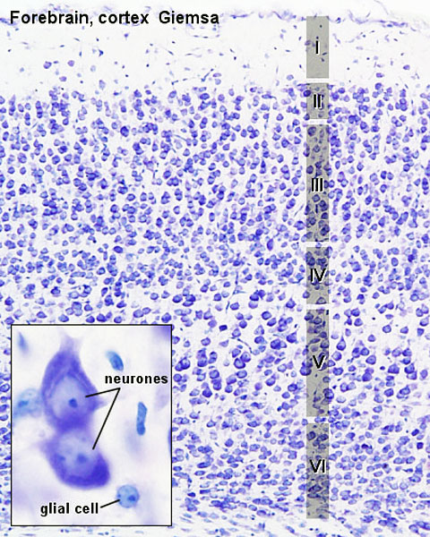 | 480 × 600 (125 KB) | Z8600021 (talk | contribs) |
You cannot overwrite this file.
File usage
The following 3 pages use this file: