File:Blood vessel wall cartoon.jpg: Difference between revisions
From Embryology
No edit summary |
mNo edit summary |
||
| Line 1: | Line 1: | ||
==Blood vessel wall | ==Blood Vessel Wall== | ||
===tunica intima=== | |||
* delimits the vessel wall towards the lumen | |||
* endothelial lining (typically simple, squamous) and associated connective tissue. | |||
* beneath the connective tissue, '''internal elastic lamina''' | |||
** forms the limit of the tunica intima | |||
===tunica media=== | |||
* formed by a layer of circumferential smooth muscle and variable amounts of connective tissue. | |||
* located beneath the smooth muscle a second layer of elastic fibers, '''external elastic lamina''' | |||
** forms the limit of the tunica media | |||
===tunica adventitia=== | |||
* consist mainly of connective tissue fibres. | |||
* blends with the connective tissue surrounding the vessel. | |||
** definition of the outer limit of the tunica adventitia is somewhat arbitrary. | |||
{{Blue Histology}} | {{Blue Histology}} | ||
[[Category:Cardiovascular]] [[Category:Cartoon]] | |||
Revision as of 08:52, 9 May 2013
Blood Vessel Wall
tunica intima
- delimits the vessel wall towards the lumen
- endothelial lining (typically simple, squamous) and associated connective tissue.
- beneath the connective tissue, internal elastic lamina
- forms the limit of the tunica intima
tunica media
- formed by a layer of circumferential smooth muscle and variable amounts of connective tissue.
- located beneath the smooth muscle a second layer of elastic fibers, external elastic lamina
- forms the limit of the tunica media
tunica adventitia
- consist mainly of connective tissue fibres.
- blends with the connective tissue surrounding the vessel.
- definition of the outer limit of the tunica adventitia is somewhat arbitrary.
Links: Histology | Histology Stains | Blue Histology images copyright Lutz Slomianka 1998-2009. The literary and artistic works on the original Blue Histology website may be reproduced, adapted, published and distributed for non-commercial purposes. See also the page Histology Stains.
Cite this page: Hill, M.A. (2024, May 19) Embryology Blood vessel wall cartoon.jpg. Retrieved from https://embryology.med.unsw.edu.au/embryology/index.php/File:Blood_vessel_wall_cartoon.jpg
- © Dr Mark Hill 2024, UNSW Embryology ISBN: 978 0 7334 2609 4 - UNSW CRICOS Provider Code No. 00098G
File history
Click on a date/time to view the file as it appeared at that time.
| Date/Time | Thumbnail | Dimensions | User | Comment | |
|---|---|---|---|---|---|
| current | 17:21, 7 July 2012 | 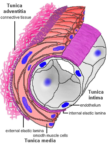 | 450 × 600 (71 KB) | Z8600021 (talk | contribs) | {{Blue Histology}} |
You cannot overwrite this file.
File usage
The following 3 pages use this file: