File:Bat-craniofacial development.jpg

Original file (600 × 720 pixels, file size: 140 KB, MIME type: image/jpeg)
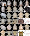
Craniofacial development of M. schreibersii fuliginosus, H. armiger and H. pratti.
(A1-10) M. schreibersii fuliginosus: (A1) Lateral view of left side of Stage 13 head;
(A2-10) Face-on views of Stage 14 (A2), Stage 15 (A3), Stage 16 (A4), Stage 17 (A5), Stage 18 (A6), Stage 20 (A7), Stage 23 (A8), fetal stage (A9), and adult (A10) heads. (B1-10) H. armiger: (B1) Lateral view of left side of Stage 11 head; (A2-9)
Face-on views of Stage 14 (B2), Stage 17 (B3), Stage 18 (B4), Stage 19 (B5), Stage 20 (B6), Stage 22 (B7), fetal stage (B8), and adult (B9) heads;
(B10) A diagram of an adult H. armiger's face illustrating nose-leaves. (C1-10) H. pratti: (C1) Lateral view of left side of Stage 11 head; (C2-9) Face-on views of Stage 14 (C2), Stage 15 (C3), Stage 16 (C4), Stage 19 (C5), Stage 20 (C6), Stage 22 (C7), fetal stage (C8), and adult (C9) heads;
(C10) A diagram of an adult H. pratti's face illustrating nose-leaves.
ah, auditory hillocks; at, antitragus; cc, chin cleft; el, eyelid; ep, eyelid primordium; fd, fold; ff, the 4th fold; fs, frontal sac; fsp, frontal sac primordium; ga, glossopharyngeal arch; ha, hyoid arch; hr, hair; lp, lens placode; lv, lens vesicle; md, mandible; mx, maxilla; ma, mandibular arch; mnl, main nose-leaf; nl, nose-leaf; nlp, nose-leaf primordium; np, nasal pit; nr, naris; og, oral groove; ope, optic evagination; opc, optic cup; otv, otic vesicle; pi, pinna; pig, pigment; pr, pigmented retina; pt, protuberance; sf, the second fold; sh, shield; tg, tragus; to, tongue; tp, tooth primordium; vf, vibrissal follicles.
Bar = 200 μm in A1-5, B1-2 and C1-3; bar = 1 mm in A6-9, B3-8 and C4-8; bar = 2 mm in A10, B9 and C9.
Original File Name: 1471-213X-10-10-3.jpg
BMC Dev Biol. 2010; 10: 10.
Published online 2010 January 21. doi: 10.1186/1471-213X-10-10. Copyright ©2010 Wang et al; licensee BioMed Central Ltd.
<pubmed>20092640</pubmed>| PMC: 2824742
This is an Open Access article distributed under the terms of the Creative Commons Attribution License (http://creativecommons.org/licenses/by/2.0), which permits unrestricted use, distribution, and reproduction in any medium, provided the original work is properly cited.
File history
Click on a date/time to view the file as it appeared at that time.
| Date/Time | Thumbnail | Dimensions | User | Comment | |
|---|---|---|---|---|---|
| current | 01:03, 29 March 2010 |  | 600 × 720 (140 KB) | S8600021 (talk | contribs) | Craniofacial development of M. schreibersii fuliginosus, H. armiger and H. pratti. (A1-10) M. schreibersii fuliginosus: (A1) Lateral view of left side of Stage 13 head; (A2-10) Face-on views of Stage 14 (A2), Stage 15 (A3), Stage 16 (A4), Stage 17 (A5), |
You cannot overwrite this file.
File usage
The following page uses this file: