File:Bartelmez1923 fig06.jpg

Original file (1,280 × 809 pixels, file size: 136 KB, MIME type: image/jpeg)
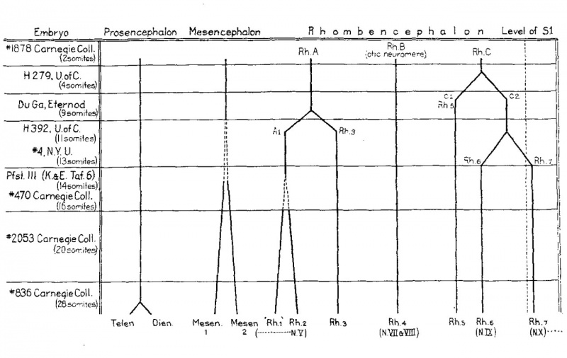
Fig. 6 The embryos here included are selected as typical of definite stages in the differentiation of the brain segments
In the horizontal columns the segments present in each embryo are shown. The bifurcations of the heavy lines indicating the individual segments represent divisions of the original segments. This division is preceded in each case by localized growth at that particular level. Where this is gradual it is shown by the gradually diverging broken lines.
| Historic Disclaimer - information about historic embryology pages |
|---|
 |
Reference
Bartelmez GW. The subdivisions of the neural folds in man. (1923) J. Comp. Neural., 35: 231-247.
Cite this page: Hill, M.A. (2024, April 27) Embryology Bartelmez1923 fig06.jpg. Retrieved from https://embryology.med.unsw.edu.au/embryology/index.php/File:Bartelmez1923_fig06.jpg
- © Dr Mark Hill 2024, UNSW Embryology ISBN: 978 0 7334 2609 4 - UNSW CRICOS Provider Code No. 00098G
File history
Click on a date/time to view the file as it appeared at that time.
| Date/Time | Thumbnail | Dimensions | User | Comment | |
|---|---|---|---|---|---|
| current | 23:06, 7 June 2016 | | 1,280 × 809 (136 KB) | Z8600021 (talk | contribs) |
You cannot overwrite this file.
File usage
The following page uses this file: