File:Baileytable02.jpg: Difference between revisions
No edit summary |
No edit summary |
||
| Line 1: | Line 1: | ||
The accompanying table indicates the types of development in the different bones of the head skeleton | ==The accompanying table indicates the types of development in the different bones of the head skeleton== | ||
Bones Of Intracartilaginous Origin | Bones Of Intracartilaginous Origin | ||
Revision as of 12:27, 18 January 2011
The accompanying table indicates the types of development in the different bones of the head skeleton
Bones Of Intracartilaginous Origin
Of Intramembranous Origin
Derived from Visceral Arches
Occipitale.
Pars basilaris. Pars lateralis. Squama occipitalis below sup. nuchal line.
Squama occipitalis above sup. nuchal line.
Temporale.
Pars mastoidea. Pars petrosa, with essus sty oideus.
proc
Pars tympanica. Squama tempo ralis.
Processus styloideus (sea arch).
Sphenoidale.
Corpus.
Ala parva.
Ala magna.
Hamulus pterygoideus.
Processus pterygoideus, except hamulus pterygoideus.
Ethmoidale.
Crista galli. Lamina cribrosa. Lamina perpendicularis. Labyrinthus ethmoidalis.
Vomer.
Vomer.
Parietale.
Parietale.
Frontale.
Frontale.
Lacrimale.
Lacrimale.
Nasale.
Nasale.
Zygoma.
Zygoma.
Maxilla.
Maxilla, with incisivum.
Maxilla,except incisivum( ?) (first arch).
Palatinum.
Palatinum.
Palatinum.
Mandibula.
Processus condyloideus,
tip of. Processus coronoideus,
tip of. Corpus, distal end of.
Processus condyloideus, except tip.
Processus coronoideus, except tip.
Corpus, except distal end.
Ramus.
Mandibula (first arch).
Hyoideum.
Hvoideum
Cornu majus (third arch). Cornu minus (second arch). Corpus (third arch).
Ossicula auditus.
Incus.
Malleus.
Stapes, except basis (?).
Basis stapedis.
Incus (first arch). Malleus (first arch). Stapes, except basis (second arch).
- Text-Book of Embryology: Germ cells | Maturation | Fertilization | Amphioxus | Frog | Chick | Mammalian | External body form | Connective tissues and skeletal | Vascular | Muscular | Alimentary tube and organs | Respiratory | Coelom, Diaphragm and Mesenteries | Urogenital | Integumentary | Nervous System | Special Sense | Foetal Membranes | Teratogenesis | Gallery of All Figures
| Historic Disclaimer - information about historic embryology pages |
|---|
 |
Reference
Bailey FR. and Miller AM. Text-Book of Embryology (1921) New York: William Wood and Co.
Cite this page: Hill, M.A. (2024, April 26) Embryology Baileytable02.jpg. Retrieved from https://embryology.med.unsw.edu.au/embryology/index.php/File:Baileytable02.jpg
- © Dr Mark Hill 2024, UNSW Embryology ISBN: 978 0 7334 2609 4 - UNSW CRICOS Provider Code No. 00098G
File history
Click on a date/time to view the file as it appeared at that time.
| Date/Time | Thumbnail | Dimensions | User | Comment | |
|---|---|---|---|---|---|
| current | 12:25, 18 January 2011 | 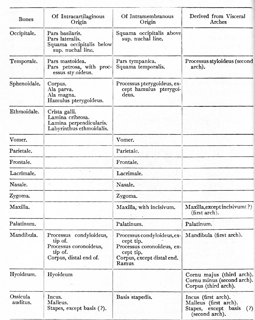 | 884 × 1,109 (182 KB) | S8600021 (talk | contribs) | The accompanying table indicates the types of development in the different bones of the head skeleton. {{Template:Bailey 1921 Figures}} Category:Human Category:Bone Category:Head |
You cannot overwrite this file.
File usage
The following 2 pages use this file: