File:Bailey322.jpg: Difference between revisions
({{Template:Bailey 1921 Figures}} Category:Human Category:Renal) |
No edit summary |
||
| Line 1: | Line 1: | ||
==Fig. 322. From a model of the cloaca and the surrounding structures in a human embryo of 6.5 mm== | |||
Keibel. | |||
The portion of the gut immediately caudal to the attachment of the allantoic duct becomes dilated to form the cloaca which at first is a blind sac, its cavity being separated from the outer surface of the embryo by the cloacal membrane (Fig. 322). | |||
The openings of the mesonephric ducts, which primarily were situated in the lateral cloacal wall (p. 359), are situated after the separation in the dorso-lateral wall of the urogenital sinus (compare Figs. 322, 323, 324). | |||
:Link: [[Book_-_Text-Book_of_Embryology_15#Fig322|Figure in Text]] | |||
{{Template:Bailey 1921 Figures}} | {{Template:Bailey 1921 Figures}} | ||
[[Category:Human]] [[Category:Renal]] | [[Category:Human]] [[Category:Renal]] | ||
Revision as of 18:11, 19 September 2011
Fig. 322. From a model of the cloaca and the surrounding structures in a human embryo of 6.5 mm
Keibel.
The portion of the gut immediately caudal to the attachment of the allantoic duct becomes dilated to form the cloaca which at first is a blind sac, its cavity being separated from the outer surface of the embryo by the cloacal membrane (Fig. 322).
The openings of the mesonephric ducts, which primarily were situated in the lateral cloacal wall (p. 359), are situated after the separation in the dorso-lateral wall of the urogenital sinus (compare Figs. 322, 323, 324).
- Link: Figure in Text
- Text-Book of Embryology: Germ cells | Maturation | Fertilization | Amphioxus | Frog | Chick | Mammalian | External body form | Connective tissues and skeletal | Vascular | Muscular | Alimentary tube and organs | Respiratory | Coelom, Diaphragm and Mesenteries | Urogenital | Integumentary | Nervous System | Special Sense | Foetal Membranes | Teratogenesis | Gallery of All Figures
| Historic Disclaimer - information about historic embryology pages |
|---|
 |
Reference
Bailey FR. and Miller AM. Text-Book of Embryology (1921) New York: William Wood and Co.
Cite this page: Hill, M.A. (2024, May 4) Embryology Bailey322.jpg. Retrieved from https://embryology.med.unsw.edu.au/embryology/index.php/File:Bailey322.jpg
- © Dr Mark Hill 2024, UNSW Embryology ISBN: 978 0 7334 2609 4 - UNSW CRICOS Provider Code No. 00098G
File history
Click on a date/time to view the file as it appeared at that time.
| Date/Time | Thumbnail | Dimensions | User | Comment | |
|---|---|---|---|---|---|
| current | 12:00, 25 January 2011 | 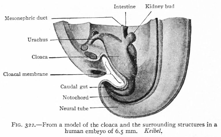 | 766 × 476 (62 KB) | S8600021 (talk | contribs) | {{Template:Bailey 1921 Figures}} Category:Human Category:Renal |
You cannot overwrite this file.
File usage
The following 3 pages use this file: