File:Bailey320.jpg
Original file (869 × 582 pixels, file size: 75 KB, MIME type: image/jpeg)
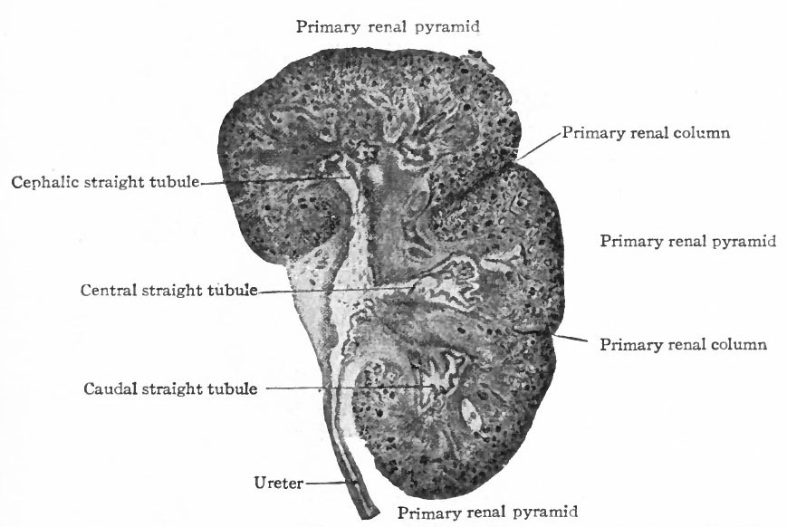
Fig. 320. Frontal section of the kidney of a human foetus of 3.75 months (10 cm)
Hauch.
The Renal Pyramids and Renal Columns
The tubules arising from the four primary evaginations of the renal pelvis together form four distinct groups or primary renal (Malpighian) pyramids one cephalic, one caudal, and two central. The central pyramids are crowded in between the end pyramids, (cephalic and caudal) and do not develop as rapidly as the latter which soon bend around toward the ureter, thus resulting in the formation of the convex side of the kidney and a depression or hilus opposite (compare Figs. 314 and 320).
Between these four pyramids the mesenchyme remains for some time as rather distinct septa, forming the primary renal columns (columns of Bertini) which are marked by corresponding depressions on the surface of the kidney and extend to the renal pelvis. The four primary pyramids may be considered as lobes (Fig. 320). It should also be stated that the parts of the tubules derived from the mesenchyme form the bases of the renal pyramids.
--Mark Hill 14:49, 27 May 2011 (EST) Fetus CRL 10cm should be about fertilization age 13 weeks or a gestational age (LMP) 15 weeks.
- Link: Figure in Text
- Text-Book of Embryology: Germ cells | Maturation | Fertilization | Amphioxus | Frog | Chick | Mammalian | External body form | Connective tissues and skeletal | Vascular | Muscular | Alimentary tube and organs | Respiratory | Coelom, Diaphragm and Mesenteries | Urogenital | Integumentary | Nervous System | Special Sense | Foetal Membranes | Teratogenesis | Gallery of All Figures
| Historic Disclaimer - information about historic embryology pages |
|---|
 |
Reference
Bailey FR. and Miller AM. Text-Book of Embryology (1921) New York: William Wood and Co.
Cite this page: Hill, M.A. (2024, April 27) Embryology Bailey320.jpg. Retrieved from https://embryology.med.unsw.edu.au/embryology/index.php/File:Bailey320.jpg
- © Dr Mark Hill 2024, UNSW Embryology ISBN: 978 0 7334 2609 4 - UNSW CRICOS Provider Code No. 00098G
File history
Click on a date/time to view the file as it appeared at that time.
| Date/Time | Thumbnail | Dimensions | User | Comment | |
|---|---|---|---|---|---|
| current | 11:59, 25 January 2011 | | 869 × 582 (75 KB) | S8600021 (talk | contribs) | {{Template:Bailey 1921 Figures}} Category:Human Category:Renal |
You cannot overwrite this file.
File usage
The following 2 pages use this file: