File:Bailey159.jpg
From Embryology
Size of this preview: 624 × 599 pixels. Other resolution: 933 × 896 pixels.
Original file (933 × 896 pixels, file size: 238 KB, MIME type: image/jpeg)
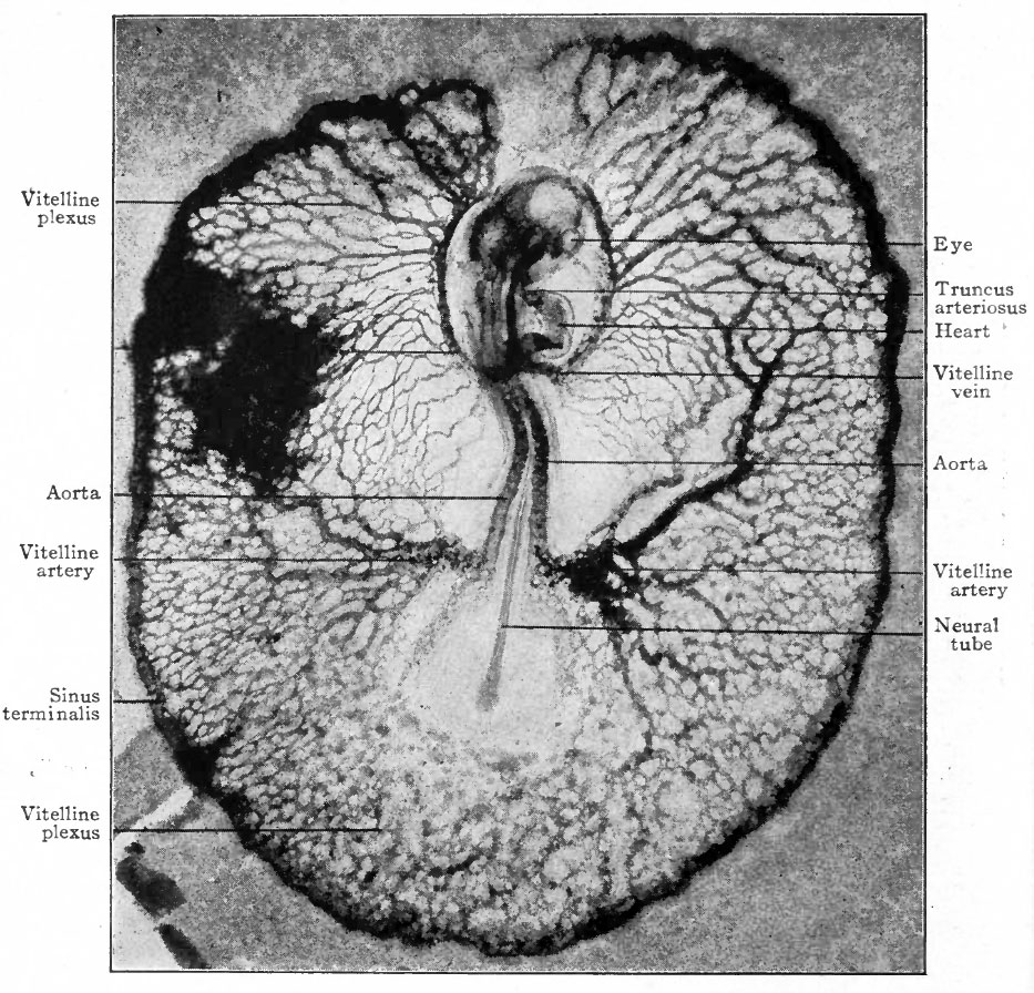
Fig. 159. Dorsal surface view of chick embryo with 18 segments, including the area vasculosa
Photograph, X 15.
The blood vessels were injected with India ink, the dark blotch in the upper left corner indicating some ink which escaped during the injection.
- Text-Book of Embryology: Germ cells | Maturation | Fertilization | Amphioxus | Frog | Chick | Mammalian | External body form | Connective tissues and skeletal | Vascular | Muscular | Alimentary tube and organs | Respiratory | Coelom, Diaphragm and Mesenteries | Urogenital | Integumentary | Nervous System | Special Sense | Foetal Membranes | Teratogenesis | Gallery of All Figures
| Historic Disclaimer - information about historic embryology pages |
|---|
 |
Reference
Bailey FR. and Miller AM. Text-Book of Embryology (1921) New York: William Wood and Co.
Cite this page: Hill, M.A. (2024, April 27) Embryology Bailey159.jpg. Retrieved from https://embryology.med.unsw.edu.au/embryology/index.php/File:Bailey159.jpg
- © Dr Mark Hill 2024, UNSW Embryology ISBN: 978 0 7334 2609 4 - UNSW CRICOS Provider Code No. 00098G
File history
Click on a date/time to view the file as it appeared at that time.
| Date/Time | Thumbnail | Dimensions | User | Comment | |
|---|---|---|---|---|---|
| current | 14:14, 18 January 2011 | | 933 × 896 (238 KB) | S8600021 (talk | contribs) | ==Fig. 159. Dorsal surface view of chick embryo with 18 segments, including the area vasculosa== Photograph, X 15. The blood vessels were injected with India ink, the dark blotch in the upper left corner indicating some ink which escaped during the inj |
You cannot overwrite this file.
File usage
The following 2 pages use this file: