File:Arthrogryposis.jpg
Arthrogryposis.jpg (800 × 503 pixels, file size: 39 KB, MIME type: image/jpeg)
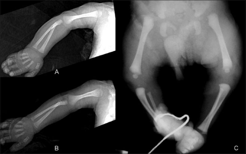
Arthrogryposis
Arthrogryposis associated with more than 300 different disorders and result from a range of developmental processes (neurogenic, myopathic, or connective tissue disorder) or environmental effects (intrauterine compression, maternal disease, teratogenic exposure or vascular insult). These x-rays show examples of limb abnormalities, postnatally visible externally and analysed by x-ray. See also recent review PMID 21851751.
- A - Frontal radiograph of the elbow show extended elbows, pronated forearms, and flexed wrists and fingers in a baby with arthrogryposis.
- B - Lateral radiograph of the elbow show extended elbows, pronated forearms, and flexed wrists and fingers in a baby with arthrogryposis.
- C - Frontal radiograph of the lower limbs of another baby with arthrogryposis shows bilateral hip dislocations and club feet.
- Links: Limb Abnormalities | Limb Development
Original File Name: Figure 15 IJRI-20-174-g015.jpg http://www.ncbi.nlm.nih.gov/pmc/articles/PMC2963757/figure/F0015/
Reference
<pubmed>21042439</pubmed>| PMC2963757
This is an open-access article distributed under the terms of the Creative Commons Attribution License, which permits unrestricted use, distribution, and reproduction in any medium, provided the original work is properly cited.
File history
Click on a date/time to view the file as it appeared at that time.
| Date/Time | Thumbnail | Dimensions | User | Comment | |
|---|---|---|---|---|---|
| current | 22:45, 2 May 2011 | | 800 × 503 (39 KB) | S8600021 (talk | contribs) | ==Arthrogryposis== Frontal (A) and lateral (B) radiographs of the elbow show extended elbows, pronated forearms, and flexed wrists and fingers in a baby with arthrogryposis. (C) Frontal radiograph of the lower limbs of another baby with arthrogryposis |
You cannot overwrite this file.
File usage
The following page uses this file: