File:Anson1934 fig01-8.jpg
Original file (1,337 × 888 pixels, file size: 126 KB, MIME type: image/jpeg)
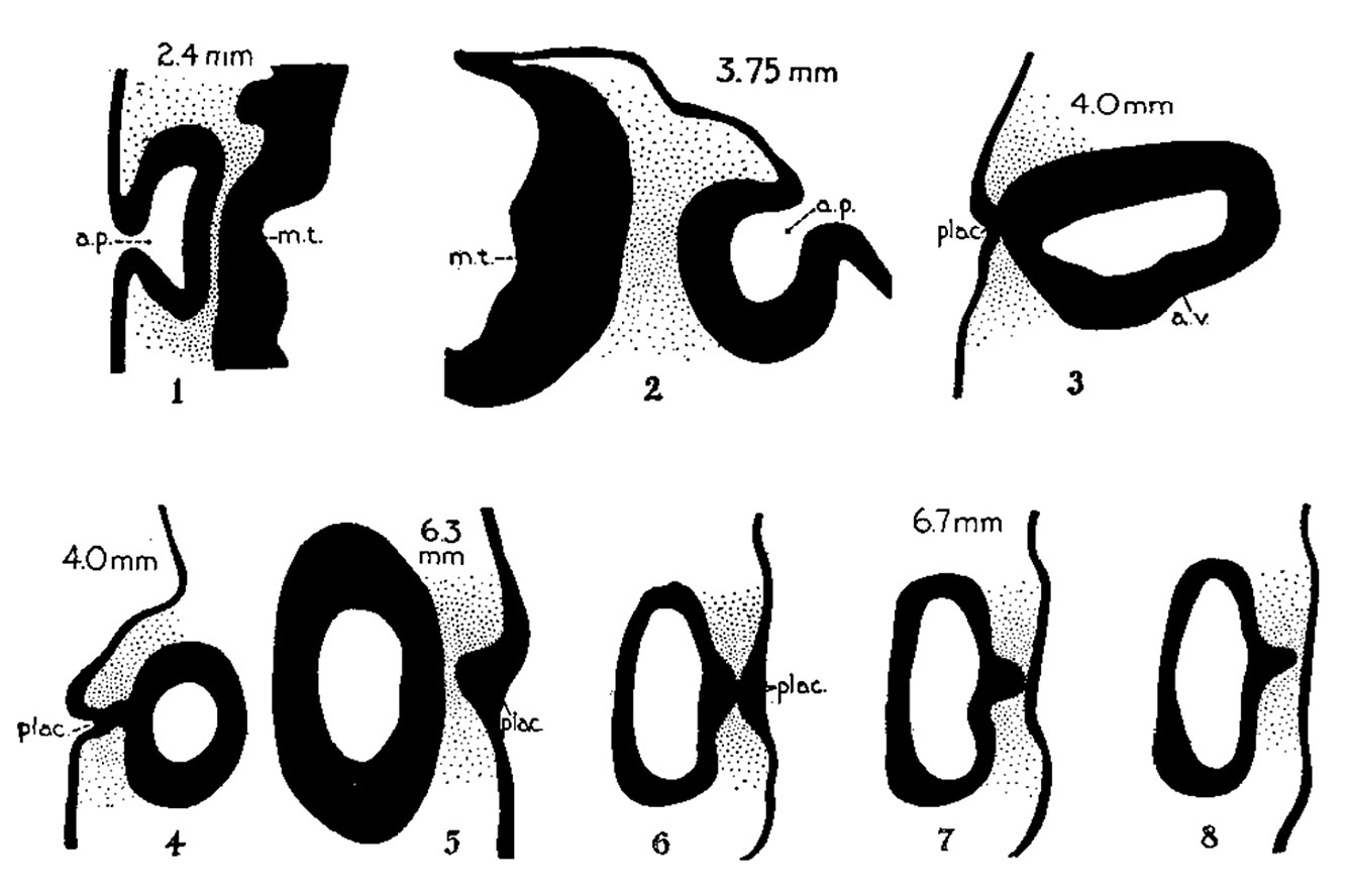
Figs. 1 to 8. Auditory Region - Transverse sections of embryos in the Harvard Embryological Collection
Harvard Embryological Collection
Dorsal aspect downward (except in 3 ) . x 80.
|
Abbreviations
a.p., auditory pit a.v., auditory vesicle (otocyst) c.d., cochlear duct coch., cochlear part of vesicle e.ap., endolymphatic appendage e.d., endolymphatic duct e.pr., eiidolymphatic projeetion e.s., endolymphatic sac ep., epidermis l.s.d., lateral semicircular duct m.t., medullary tube plac., placodal stalk sacc., saccule s.s.d., superior semicircular duct utr., utricle vest., vesibular part of vesicle |
| Historic Disclaimer - information about historic embryology pages |
|---|
 |
- Otic Vesicle Links: Fig. 1-8 | Plate 1 | 9 | 10 | 11 | 12 | 13 | 14 | Plate 2 | 15 | 16 | 17 | 18 | 19 | Anson 1934 | Harvard Collection | Inner Ear | Hearing
Reference
Anson BJ. and Black WT. The early relation of the auditory vesicle to the ectoderm in human embryos. (1934) Anat. Rec. 58: 127-137.
Cite this page: Hill, M.A. (2024, April 27) Embryology Anson1934 fig01-8.jpg. Retrieved from https://embryology.med.unsw.edu.au/embryology/index.php/File:Anson1934_fig01-8.jpg
- © Dr Mark Hill 2024, UNSW Embryology ISBN: 978 0 7334 2609 4 - UNSW CRICOS Provider Code No. 00098G
File history
Click on a date/time to view the file as it appeared at that time.
| Date/Time | Thumbnail | Dimensions | User | Comment | |
|---|---|---|---|---|---|
| current | 13:52, 23 July 2015 | | 1,337 × 888 (126 KB) | Z8600021 (talk | contribs) | |
| 13:50, 23 July 2015 |  | 1,800 × 1,556 (384 KB) | Z8600021 (talk | contribs) |
You cannot overwrite this file.
File usage
The following 2 pages use this file: