File:Aneuploidy model based on fragmentation 1.jpg
Original file (946 × 907 pixels, file size: 154 KB, MIME type: image/jpeg)
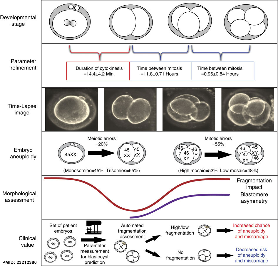
Embryonic development was monitored by time-lapse imaging from the one- to four-cell stage followed by assessment of chromosomal composition of each blastomere in the imaged embryos. We observed refinement of diagnostic non-invasive cell cycle parameters, determined the correlation with meiotic (monosomies and trisomies) and mitotic (high and low mosaic) errors and demonstrated an association between the cell cycle parameters and embryo morphology (fragmentation and blastomere asymmetry). We also suggest clinical value of parameter analysis with and without automated fragmentation assessment.
Above text from figure legend and Supplementary refers to the original article supplementary information.
- Links: Abnormal Development - Genetic | Trisomy 21 | Zygote | Morula
Reference
<pubmed>23212380</pubmed>| Nat Commun.
Copyright
http://creativecommons.org/licenses/by-nc-sa/3.0/
Figure 7. Ncomms2249-f7.jpg Original figure altered in size and labelling.
File history
Click on a date/time to view the file as it appeared at that time.
| Date/Time | Thumbnail | Dimensions | User | Comment | |
|---|---|---|---|---|---|
| current | 09:54, 12 January 2015 | | 946 × 907 (154 KB) | Z8600021 (talk | contribs) |
You cannot overwrite this file.
File usage
The following page uses this file: