File:Adrenal histology 004.jpg: Difference between revisions
From Embryology
(uploaded a new version of "File:Adrenal histology 004.jpg") |
mNo edit summary |
||
| (4 intermediate revisions by the same user not shown) | |||
| Line 1: | Line 1: | ||
==Adrenal Histology - Zona Glomerulosa and Fasciculata== | ==Adrenal Histology - Zona Glomerulosa and Fasciculata== | ||
Adrenal gland, monkey - {{HE}} | |||
===Zona Glomerulosa=== | |||
(Latin, ''glomus'' = "ball") | |||
* most superficial layer of the adrenal cortex. | |||
* beneath the capsule. | |||
* ovoid cells arranged in clusters or arches. | |||
=== Zona Fasciculata=== | |||
* middle zone of the adrenal cortex. | |||
* beneath the zona glomerulosa. | |||
* cells arranged into "fascicles" or bundles. | |||
* produces glucocorticoids (cortisol) that regulates glucose metabolism. | |||
{{Adrenal Histology}} | {{Adrenal Histology}} | ||
[[Category:Adrenal]] [[Category:Endocrine]] [[Category:Histology]] | [[Category:Adrenal]] [[Category:Endocrine]] [[Category:Histology]] | ||
Latest revision as of 15:05, 11 July 2016
Adrenal Histology - Zona Glomerulosa and Fasciculata
Adrenal gland, monkey - (Stain - Haematoxylin Eosin)
Zona Glomerulosa
(Latin, glomus = "ball")
- most superficial layer of the adrenal cortex.
- beneath the capsule.
- ovoid cells arranged in clusters or arches.
Zona Fasciculata
- middle zone of the adrenal cortex.
- beneath the zona glomerulosa.
- cells arranged into "fascicles" or bundles.
- produces glucocorticoids (cortisol) that regulates glucose metabolism.
- Adrenal Histology: Cortex and Medulla | Unlabelled Overview | Cortical Zones | Zona Glomerulosa and Fasciculata | Zona Glomerulosa | Zona Fasciculata | Zona Reticularis and Medulla | Zona Reticularis | Medulla | Fetal Cortex | Developing Adult Cortex | BGD - Endocrine Histology | Histology Stains | Adrenal Development
Links: Histology | Histology Stains | Blue Histology images copyright Lutz Slomianka 1998-2009. The literary and artistic works on the original Blue Histology website may be reproduced, adapted, published and distributed for non-commercial purposes. See also the page Histology Stains.
Cite this page: Hill, M.A. (2024, May 20) Embryology Adrenal histology 004.jpg. Retrieved from https://embryology.med.unsw.edu.au/embryology/index.php/File:Adrenal_histology_004.jpg
- © Dr Mark Hill 2024, UNSW Embryology ISBN: 978 0 7334 2609 4 - UNSW CRICOS Provider Code No. 00098G
File history
Click on a date/time to view the file as it appeared at that time.
| Date/Time | Thumbnail | Dimensions | User | Comment | |
|---|---|---|---|---|---|
| current | 14:46, 12 May 2012 | 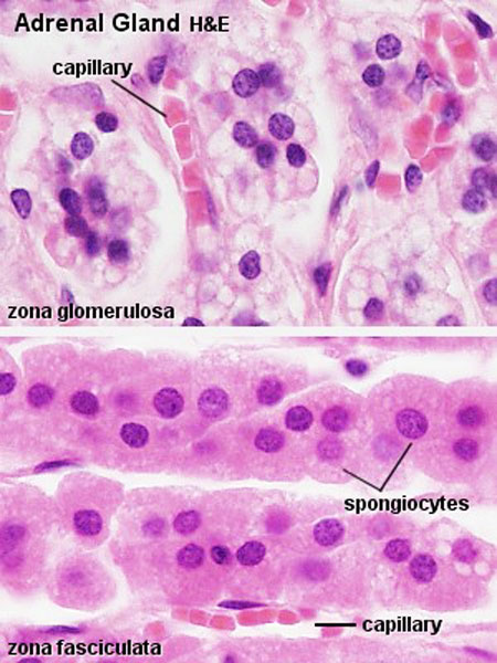 | 450 × 600 (71 KB) | Z8600021 (talk | contribs) | |
| 13:08, 28 September 2010 |  | 300 × 400 (47 KB) | S8600021 (talk | contribs) | ==Adrenal Gland Histology== Adrenal gland, monkey - H&E Original file name: Adr042he.jpg Adrenal gland histology 01.jpg {{Template:Blue Histology}} Category:Adrenal Category:Endocrine Category:Histology |
You cannot overwrite this file.
File usage
The following 4 pages use this file: