File:Mall1897 fig52.jpg

Original file (907 × 1,000 pixels, file size: 113 KB, MIME type: image/jpeg)
Fig. 52. Embryo No. XXII within the Ovum
Embryo No. XXII (22)
Enlarged 3 diameters. The villi of chorion are outlined on one side of the ovum only. The umbilical vesicle, uv, has become shifted around to the dorsal and right side of the embryo. The outline is made from a photograph, and is correct in detail with the exception of the attachment of the chord to the chorion. This in reality attaches itself to the chorion immediately to the right of the embryo.
Reference
Mall FP. Development of the human coelom. (1897) J Morphol. 12: 395-453.
Cite this page: Hill, M.A. (2026, April 18) Embryology Mall1897 fig52.jpg. Retrieved from https://embryology.med.unsw.edu.au/embryology/index.php/File:Mall1897_fig52.jpg
- © Dr Mark Hill 2026, UNSW Embryology ISBN: 978 0 7334 2609 4 - UNSW CRICOS Provider Code No. 00098G
File history
Yi efo/eka'e gwa ebo wo le nyangagi wuncin ye kamina wunga tinya nan
| Gwalagizhi | Nyangagi | Dimensions | User | Comment | |
|---|---|---|---|---|---|
| current | 15:39, 12 September 2017 | | 907 × 1,000 (113 KB) | Z8600021 (talk | contribs) | |
| 15:38, 12 September 2017 | 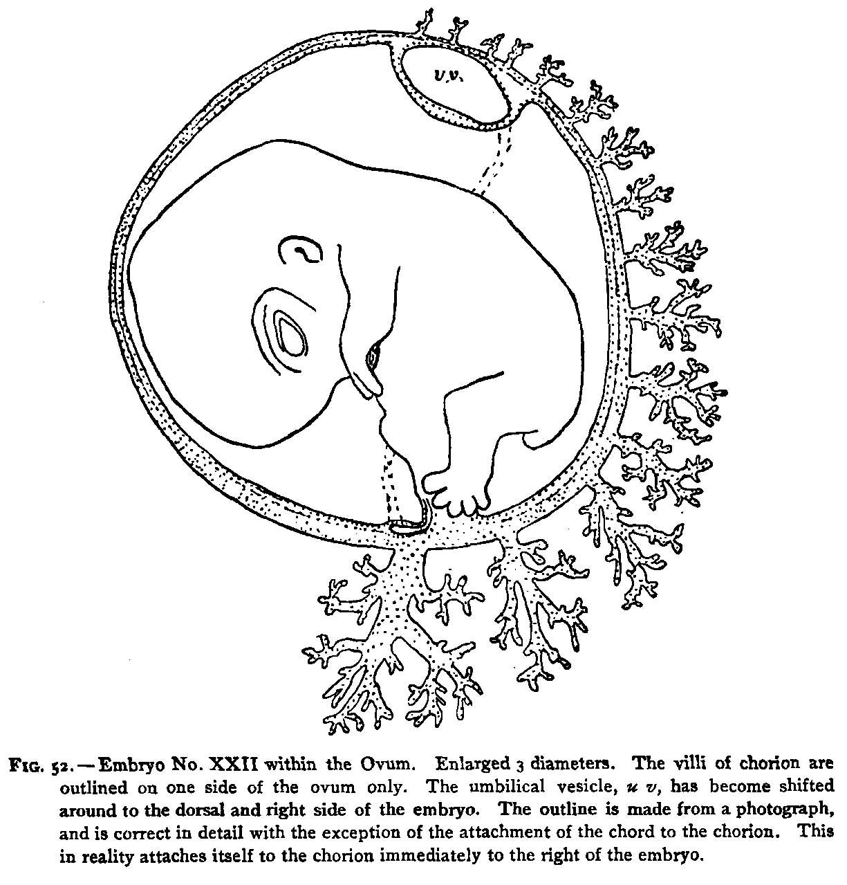 | 1,225 × 1,279 (197 KB) | Z8600021 (talk | contribs) | {{Ref-Mall1897}} |
You cannot overwrite this file.
File usage
The following page uses this file: