Category:Pig: Difference between revisions
From Embryology
mNo edit summary |
mNo edit summary |
||
| Line 7: | Line 7: | ||
Embryology Main Page - [[Pig Development|'''Pig Development''']] | |||
{{Pig}} | |||
[[Category:Animal Development]] | [[Category:Animal Development]] | ||
Revision as of 18:24, 15 December 2017
This Embryology category shows pages and media that relates to pig development.
The pig (Sus scrofa) developmental model is studied extensively due to the commercial applications of pigs for meat production and for health issues such as obesity, cardiovascular disease, and organ transplantation (xenotransplantation).
Historically, it represented an easily accessible animal embryo that could be used to model human development.
Embryology Main Page - Pig Development
Pages in category 'Pig'
The following 179 pages are in this category, out of 179 total.
B
- Book - A Laboratory Manual and Text-book of Embryology 5
- Book - A Laboratory Manual and Text-book of Embryology 6
- Book - A Laboratory Outline of Embryology 1923
- Book - A Laboratory Text-Book of Embryology 4 (1903)
- Book - Contributions to Embryology Carnegie Institution No.100
- Book - Contributions to Embryology Carnegie Institution No.54
- Book - Contributions to Embryology Carnegie Institution No.60
- Book - Developmental Anatomy 1924-17
- Book - Embryology of the Pig
- Book - Embryology of the Pig 1
- Book - Embryology of the Pig 10
- Book - Embryology of the Pig 11
- Book - Embryology of the Pig 12
- Book - Embryology of the Pig 13
- Book - Embryology of the Pig 14
- Book - Embryology of the Pig 2
- Book - Embryology of the Pig 3
- Book - Embryology of the Pig 4
- Book - Embryology of the Pig 5
- Book - Embryology of the Pig 6
- Book - Embryology of the Pig 7
- Book - Embryology of the Pig 8
- Book - Embryology of the Pig 9
- Book - Normal Plates of the Development of Vertebrates 1
H
P
- Paper - Cyclic changes in the ovaries and uterus of swine and their relations to the mechanism of implantation (1921)
- Paper - Development and homology of the mammalian cerebellar fissures 2
- Paper - Development and vascularization of the testis (1906)
- Paper - Development of the Wolffian body in Sus Scrofa Domesticus
- Paper - Double ureters in human and pig embryos
- Paper - Maturation of the ovum in swine (1917)
- Paper - Meninges histogenesis and structure
- Paper - Models of the pancreas in embryos of the pig, rabbit, cat, and man (1908)
- Paper - On the development of the atrial septum and the valvular apparatus in the right atrium of the pig embryo (1916)
- Paper - On the development of the hind-brain of the pig 1
- Paper - On the development of the hind-brain of the pig 2
- Paper - On the development of the lymphatics in the stomach of the embryo pig (1921)
- Paper - On the fate of the posterior cardinal veins and their relation to the development of the vena cava and azygos in the embryo pig (1915)
- Paper - On the histogenesis of gastric glands
- Paper - On the origin of the lymphatic system from the veins and the development of the lymph hearts and thoracic duct in the pig (1902)
- Paper - On the proportions, development and attachment of the tectorial membrane (1915)
- Paper - On the relation of the head chorda to the pharyngeal epithelium in the pig embryo
- Paper - Prenatal growth of swine (1928)
- Paper - Prenatal growth of the pig
- Paper - Some features of the histogenesis of the thyreoid gland in the pig (1910)
- Paper - Some observations on the development of the vagina in the pig (1934)
- Paper - Structure and development of the pig skull
- Paper - The anatomy of a 7.8 mm pig embryo
- Paper - The beginning and development of function in the suprarenal medulla of pig embryos (1922)
- Paper - The corpus luteum of pregnancy, as it is in swine (1915)
- Paper - The development of a medial motor nucleus and an accessory abducens nucleus in the pig (1934)
- Paper - The development of the cardiac-coronary circulatory system
- Paper - The development of the cerebral ventricles in the pig (1913)
- Paper - The development of the hypoglossal ganglia of pig embryos
- Paper - The development of the lobule of the pig's liver (1919)
- Paper - The development of the principal arterial stems in the forelimb of the pig (1922)
- Paper - The development of the spiral coil in the large intestine of the pig
- Paper - The development of the sympathetic nervous system in mammals
- Paper - The development of the thymus
- Paper - The development of the thymus in the pig 1 (1915)
- Paper - The development of the thymus in the pig 2 (1915)
- Paper - The development of the veins in the limbs of rabbit embryos
- Paper - The early development of the mammalian sternum
- Paper - The early morphogenesis and histogenesis of the liver in Sus scrofa domesticus, including notes on the morphogenesis of the ventral pancreas
- Paper - The Embryonic Development of the Interstitial Cells of Leydig (1904)
- Paper - The embryonic development of the ovary and testis of the mammals (1904)
- Paper - The fate of the ultimobranchial bodies in the pig (1918)
- Paper - The gross anatomy of a 12 mm pig
- Paper - The histogenesis of the adrenal in the pig (1903)
- Paper - The histology of an hermaphrodite pig and its developmental significance (1929)
- Paper - The microscopic structure of the yolk-sac of the pig embryo (1916)
- Paper - The organ of jacobson in the horse, ox, camel and pig (1925)
- Paper - The pharyngeal pouches and their derivatives in the mammalia
- Paper - The regular occurrence of intestinal diverticula in embryos of the pig, rabbit and man
- Paper - The regular occurrence of intestinal diverticula in embryos of the pig, rabbit, and man
- Template:Patten1951 TOC
- Template:Pig
- Pig Development
- Template:Pig links
R
- Template:Ref-Angle1918
- Template:Ref-Badertscher1915a
- Template:Ref-Badertscher1915b
- Template:Ref-Badertscher1918
- Template:Ref-Badertscher1919
- Template:Ref-Bardeen1900
- Template:Ref-Baumgartner1916
- Template:Ref-Baxter1934
- Template:Ref-BaxterBoyd1938
- Template:Ref-Boyden1955
- Template:Ref-Bradley1905
- Template:Ref-Bradley1906
- Template:Ref-Brambell1929
- Template:Ref-Carey1922
- Template:Ref-Cash1921
- Template:Ref-ClarkER1915
- Template:Ref-Corner1914
- Template:Ref-Corner1915
- Template:Ref-Corner1917
- Template:Ref-Corner1919
- Template:Ref-Corner1920
- Template:Ref-Corner1921
- Template:Ref-Corner1922b
- Template:Ref-CornerAmsbaugh1917
- Template:Ref-Cunningham1916
- Template:Ref-Davis1910
- Template:Ref-Flint1906
- Template:Ref-Goldsmith1937
- Template:Ref-Hardesty1915a
- Template:Ref-Hardesty1915b
- Template:Ref-Heuser1913
- Template:Ref-HeuserStreeter1929
- Template:Ref-Hill1905
- Template:Ref-Hilton1903
- Template:Ref-HuberCurtis1913
- Template:Ref-Johnson1919
- Template:Ref-Jones1914
- Template:Ref-Jordan1916
- Template:Ref-Jordan1919
- Template:Ref-Kampmeier1912
- Template:Ref-Keibel1897
- Template:Ref-Kingsbury1909
- Template:Ref-Kirk1910
- Template:Ref-Lewis1903
- Template:Ref-LewisPapez1915
- Template:Ref-LewisThyng1908
- Template:Ref-LillieMoore1923
- Template:Ref-Lineback1916
- Template:Ref-Lowrey1911
- Template:Ref-McClendon1913
- Template:Ref-McGill1910
- Template:Ref-Minett1925
- Template:Ref-Moody1910
- Template:Ref-Morrill1916
- Template:Ref-Parker1874
- Template:Ref-Patten1920
- Template:Ref-Pohlman1919
- Template:Ref-Prentiss1910
- Template:Ref-Rand1917
- Template:Ref-Reinke1910
- Template:Ref-Sabin1902a
- Template:Ref-Sabin1904
- Template:Ref-Sabin1905
- Template:Ref-Sabin1905a
- Template:Ref-Sabin1915
- Template:Ref-Sabin1915 figures
- Template:Ref-Sabin1917
- Template:Ref-Shaner1934
- Template:Ref-Smith1909
- Template:Ref-Solomons1924
- Template:Ref-Streeter1927
- Template:Ref-Streeter1927a
- Template:Ref-Thyng1908
- Template:Ref-Thyng1911
- Template:Ref-Warwick1928
- Template:Ref-Weed1917
- Template:Ref-Weymann1922
- Template:Ref-Weysse1894
- Template:Ref-White1939
- Template:Ref-Whitehead1903
- Template:Ref-Witte1919
- Template:Ref-Woollard1922
S
Media in category 'Pig'
The following 151 files are in this category, out of 151 total.
 Allen1904 plate7.jpg 1,280 × 1,837; 704 KB
 Arey1924 fig364.jpg 1,000 × 856; 151 KB
 Arey1924 fig365.jpg 1,000 × 1,131; 178 KB
 Arey1924 fig366.jpg 1,525 × 1,091; 336 KB
 Arey1924 fig367.jpg 1,000 × 913; 220 KB
 Arey1924 fig368.jpg 1,200 × 1,162; 312 KB
 Arey1924 fig369.jpg 1,200 × 1,231; 221 KB
 Arey1924 fig370.jpg 800 × 478; 60 KB
 Arey1924 fig371.jpg 1,000 × 626; 130 KB
 Arey1924 fig372.jpg 800 × 423; 58 KB
 Arey1924 fig373.jpg 1,200 × 997; 282 KB
 Arey1924 fig374.jpg 1,200 × 987; 268 KB
 Arey1924 fig418.jpg 1,280 × 1,498; 343 KB
 Arey1924 fig419.jpg 700 × 493; 25 KB
 Bailey110.jpg 481 × 643; 110 KB
 Bailey111.jpg 916 × 567; 117 KB
 Bailey116.jpg 835 × 434; 90 KB
 Bailey124.jpg 877 × 547; 114 KB
 Bailey126.jpg 903 × 693; 168 KB
 Bailey177.jpg 943 × 873; 199 KB
 Bailey213.jpg 800 × 851; 101 KB
 Bailey214.jpg 439 × 781; 81 KB
 Bailey259.jpg 690 × 414; 95 KB
 Bailey280.jpg 866 × 581; 133 KB
 Bailey286.jpg 842 × 481; 85 KB
 Bailey290.jpg 935 × 635; 154 KB
 Bailey327.jpg 872 × 567; 89 KB
 Bailey350.jpg 925 × 640; 129 KB
 Bailey351.jpg 749 × 598; 133 KB
 Bailey383.jpg 859 × 455; 118 KB
 Bailey385.jpg 787 × 683; 165 KB
 Bailey489.jpg 517 × 617; 82 KB
 Bailey490.jpg 755 × 612; 71 KB
 Bailey503.jpg 606 × 624; 76 KB
 Baumgartner1916 plate01.jpg 1,692 × 2,228; 308 KB
 Baumgartner1916 plate02.jpg 1,613 × 2,204; 399 KB
 Borwn014.jpg 800 × 770; 91 KB
 Brown001.jpg 600 × 388; 52 KB
 Brown003.jpg 800 × 630; 85 KB
 Brown004.jpg 800 × 618; 93 KB
 Brown005.jpg 800 × 753; 99 KB
 Brown007.jpg 800 × 594; 78 KB
 Brown008.jpg 800 × 745; 112 KB
 Brown009.jpg 800 × 660; 78 KB
 Brown010.jpg 400 × 513; 21 KB
 Brown011.jpg 400 × 473; 25 KB
 Brown012.jpg 800 × 745; 105 KB
 Brown014.jpg 800 × 770; 91 KB
 Brown016.jpg 800 × 742; 124 KB
 Brown017.jpg 625 × 800; 84 KB
 Brown018.jpg 764 × 800; 85 KB
 Brown019.jpg 911 × 1,042; 146 KB
 Brown020.jpg 723 × 800; 105 KB
 Brown022.jpg 625 × 800; 93 KB
 Corner-plate01.jpg 653 × 935; 160 KB
 Corner-plate02.jpg 729 × 986; 152 KB
 Corner-plate03.jpg 744 × 960; 150 KB
 Corner-plate04.jpg 719 × 974; 193 KB
 Corner-table01.jpg 600 × 335; 36 KB
 Corner-table02.jpg 800 × 647; 107 KB
 Corner001.jpg 858 × 356; 44 KB
 Corner002.jpg 873 × 723; 88 KB
 Corner002a.jpg 848 × 307; 49 KB
 Corner002b.jpg 849 × 328; 33 KB
 Corner1920 fig01.jpg 1,000 × 606; 159 KB
 Corner1920 fig02-05.jpg 1,000 × 671; 216 KB
 Corner1920 Plate 1.jpg 756 × 1,000; 222 KB
 Corner1922 fig01.jpg 718 × 597; 81 KB
 Corner1922 plate01.jpg 1,560 × 2,000; 433 KB
 Corner1922 plate02.jpg 1,224 × 2,000; 526 KB
 Flint1906 plate01.jpg 1,280 × 843; 149 KB
 Flint1906 textfig01.jpg 700 × 473; 33 KB
 Foster118a.jpg 724 × 1,003; 175 KB
 Gray0976.jpg 600 × 599; 146 KB
 Johnston1907 fig020.jpg 1,280 × 1,632; 277 KB
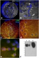
 Karyotype of parthenogenetic blastocysts.jpg 970 × 793; 121 KB
 Keibel Mall 2 005.jpg 1,000 × 916; 330 KB
 Keibel Mall 2 255.jpg 1,665 × 800; 163 KB
 Keibel Mall 2 440.jpg 1,280 × 1,256; 256 KB
 Keibel Mall 2 465.jpg 1,280 × 1,015; 272 KB
 Keibel Mall 2 512.jpg 1,000 × 1,119; 281 KB
 Keibel Mall 2 517.jpg 898 × 833; 80 KB
 Keibel Mall 2 518.jpg 996 × 833; 76 KB
 Keibel Mall 2 519.jpg 1,280 × 1,389; 271 KB
 Keibel Mall 222.jpg 684 × 543; 84 KB
 Keibel Mall 325.jpg 301 × 577; 60 KB
 Keibel Mall 326.jpg 800 × 641; 147 KB
 Keibel Mall 327.jpg 735 × 900; 152 KB
 Keibel Mall 328.jpg 800 × 774; 125 KB
 Keibel Mall 329.jpg 800 × 464; 69 KB
 Keibel1897 plate01.jpg 1,504 × 2,000; 365 KB
 Keibel1897 plate02.jpg 1,815 × 2,259; 354 KB
 Keibel1897 plate03.jpg 1,838 × 2,271; 416 KB
 Lewis1903 plate01.jpg 1,280 × 1,639; 328 KB
 Lewis1903 plate02.jpg 1,280 × 1,552; 258 KB
 Lewis1903 plate03.jpg 1,280 × 1,593; 442 KB
 Lewis1903 plate04.jpg 1,280 × 1,720; 403 KB
 LewisFT1920 fig10-13.jpg 2,348 × 3,299; 442 KB
 LewisFT1920 fig10.jpg 1,088 × 808; 46 KB
 Lineback1920 fig06-7.jpg 1,200 × 808; 177 KB
 Merkel cell EM 01.jpg 984 × 738; 209 KB
 Merkel cell EM 02.jpg 984 × 685; 166 KB
 Minot1897 001.jpg 416 × 508; 56 KB
 Minot1897 436.jpg 1,200 × 843; 146 KB
 Model capacitation-induced acrosome docking to sperm membrane.jpg 600 × 489; 73 KB
 Ovary follicle size graph.jpg 1,057 × 820; 102 KB
 Ovary oocyte size graph.jpg 1,057 × 820; 114 KB
 Patterns of ZPC Deposition in Porcine Oocytes.jpg 401 × 600; 84 KB
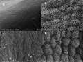 Pig - uterine epithelium SEM.jpg 800 × 598; 118 KB
 Pig blastocyst day8.jpg 500 × 505; 14 KB
 Pig development day 1-10.jpg 718 × 1,000; 112 KB
 Pig lung alveolarization.jpg 600 × 389; 38 KB
 Pig sperm capacitation 01.jpg 1,000 × 840; 204 KB
 Pig sperm capacitation 02.jpg 600 × 504; 82 KB
 Prentiss1906 fig03.jpg 1,289 × 595; 168 KB
 Prentiss1906 fig04.jpg 1,618 × 1,064; 490 KB
 Prentiss1906 fig05.jpg 1,448 × 2,283; 988 KB
 Reagan1912 fig09-15.jpg 1,581 × 1,895; 222 KB
 Reagan1912 fig16.jpg 1,548 × 1,096; 191 KB
 Reagan1912 plate03.jpg 1,435 × 1,305; 154 KB
 Rugh 063.jpg 1,200 × 755; 173 KB
 Sabin1915 plate01.jpg 2,561 × 3,250; 922 KB
 Sabin1915 plate02.jpg 2,111 × 2,788; 609 KB
 Sabin1915 plate03.jpg 2,236 × 3,033; 1.08 MB
 Sabin1915 plate04.jpg 2,023 × 2,907; 797 KB
 Sabin1915 plate05.jpg 2,861 × 2,231; 1,013 KB
 Sabin1915 plate06.jpg 2,741 × 2,269; 926 KB
 Sabin1915 plate07.jpg 2,275 × 2,920; 1.16 MB
 Sabin1915.pdf ; 6.25 MB
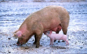
 Sow and piglet.jpg 600 × 372; 43 KB
 Streeter1906 fig07.jpg 1,254 × 591; 75 KB
 Streeter1907 fig03.jpg 545 × 666; 66 KB
 Streeter1907 fig04.jpg 572 × 565; 77 KB
 Streeter1927 plate01.jpg 1,435 × 2,000; 897 KB
 Stricht-plate01.jpg 1,156 × 1,500; 243 KB
 Stricht-plate02.jpg 1,016 × 1,261; 308 KB
 Stricht-plate03.jpg 1,023 × 1,260; 230 KB
 Stricht-plate04.jpg 1,013 × 1,251; 238 KB
 Whitehead1904 fig01.jpg 800 × 881; 90 KB
 Whitehead1904 fig02.jpg 500 × 442; 47 KB
 Whitehead1904 fig03.jpg 700 × 752; 114 KB
 Whitehead1904 fig04.jpg 800 × 669; 105 KB
 Whitehead1904 fig05.jpg 800 × 719; 124 KB
 Whitehead1904 fig06.jpg 500 × 421; 42 KB
 Woollard-plate01.jpg 744 × 1,000; 153 KB
 Woollard-plate02.jpg 788 × 1,000; 196 KB
 Woollard001.jpg 739 × 858; 142 KB
 Woollard002.jpg 699 × 873; 125 KB
 Woollard003.jpg 1,107 × 848; 159 KB
 Woollard004.jpg 1,037 × 717; 148 KB
 Woollard005.jpg 1,034 × 655; 144 KB


