Category:Immune
From Embryology
This is a direct link to pages, images and media related to immune system development.
Subcategories
This category has the following 4 subcategories, out of 4 total.
Pages in category 'Immune'
The following 82 pages are in this category, out of 82 total.
A
B
C
H
I
L
M
P
- Paper - Histogenesis and morphogenesis of the thoracic duct in the chick (1913)
- Paper - On the cervical veins and lymphatics in four human embryos, with an interpretation of anomalies on the subclavian and jugular veins in the adult (1909)
- Paper - On the development of lymphatic nodes in the pig and their relation to the lymph hearts (1905)
- Paper - Origin and development of the primitive vessels of the chick and of the pig (1917)
- Paper - Preliminary note on the differentiation of angioblasts in the living chick (1917)
- Paper - The anatomy and development of the systemic lymphatic vessels in the domestic cat
- Paper - The development of the human pharyngeal tonsil
- Paper - The development of the lymphatic system in rabbits
- Paper - The development of the mammalian spleen, with special reference to its hematopoietic activity (1921)
- Paper - The developmental significance of the mammalian pharyngeal tonsil - Cat
- Paper - The first lymph glands in rabbits and human embryos (1909)
- Paper - The morphogenesis and histogenesis of the thymus gland in man
- Paper - The parathyroid glands and the lateral thyroid in man
- Paper - The phylogenetic relations of the lymphatic and bloodvascular systems in vertebrates (1910)
- Postnatal - Vaccination
R
- Template:Ref-Clark1915b
- Template:Ref-Clark1920
- Template:Ref-ClarkEL1912
- Template:Ref-ClarkER1915
- Template:Ref-Huntington1910
- Template:Ref-Huntington1910a
- Template:Ref-Huntington1910b
- Template:Ref-Huntington1911
- Template:Ref-Kampmeier1912
- Template:Ref-Kingsbury1932
- Template:Ref-Lewis1905a
- Template:Ref-LewisFT1909
- Template:Ref-MillerA1913
- Template:Ref-Sabin1911b
- Template:Ref-Sabin1912
- Template:Ref-Snook1934a
S
Media in category 'Immune'
The following 157 files are in this category, out of 157 total.
 Adult human liver cells.jpg 900 × 1,095; 245 KB
 Adult lymphatic system cartoon.jpg 800 × 755; 110 KB
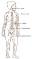 Adult lymphatic system.jpg 329 × 600; 22 KB
 Arey1924 fig201.jpg 1,200 × 1,588; 183 KB
 Aus-Imm-Handbook-2015.jpg 420 × 595; 28 KB
 B lymphocyte EM08.jpg 1,000 × 730; 111 KB
 B lymphocyte EM09.jpg 673 × 1,000; 89 KB
 B lymphocyte EM10.jpg 671 × 1,000; 92 KB
 B lymphocytes EM08-10.jpg 677 × 996; 124 KB
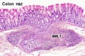 Colon MALT.jpg 500 × 333; 67 KB
 Community immunity cartoon.jpg 537 × 732; 233 KB
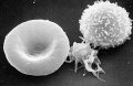 Erythrocyte and lymphocyte SEM01.jpg 800 × 522; 74 KB
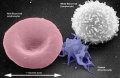 Erythrocyte and lymphocyte SEM02.jpg 800 × 522; 78 KB
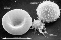 Erythrocyte and lymphocyte SEM03.jpg 800 × 522; 80 KB
 Fetal thymus weight growth graph.jpg 1,000 × 669; 52 KB
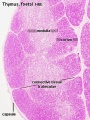 Fetal thymus.jpg 450 × 600; 122 KB
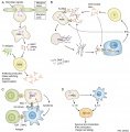
 Gastrointestinal tract intestine immune cartoon 01.jpg 728 × 1,200; 284 KB
 Gray0592.jpg 600 × 590; 55 KB
 Gray0593.jpg 550 × 437; 66 KB
 Gray0594.jpg 600 × 432; 83 KB
 Gray0595.jpg 600 × 491; 94 KB
 Gray0596.jpg 600 × 390; 71 KB
 Gray0597.jpg 700 × 496; 80 KB
 Gray0598.jpg 600 × 443; 83 KB
 Gray0599.jpg 413 × 1,000; 109 KB
 Gray0600.jpg 1,200 × 528; 100 KB
 Gray0601.jpg 1,000 × 357; 62 KB
 Gray0602.jpg 700 × 708; 132 KB
 Gray0603.jpg 800 × 480; 69 KB
 Gray0604.jpg 716 × 500; 65 KB
 Gray0605.jpg 614 × 600; 98 KB
 Gray0606.jpg 686 × 800; 93 KB
 Gray0607.jpg 800 × 623; 126 KB
 Gray0608.jpg 400 × 623; 74 KB
 Gray0609.jpg 527 × 500; 69 KB
 Gray0610.jpg 303 × 1,000; 81 KB
 Gray0611.jpg 650 × 600; 123 KB
 Gray0612.jpg 800 × 631; 181 KB
 Gray0613.jpg 700 × 579; 115 KB
 Gray0614.jpg 550 × 640; 118 KB
 Gray0615.jpg 571 × 600; 101 KB
 Gray0616.jpg 626 × 600; 111 KB
 Gray0617.jpg 555 × 600; 139 KB
 Gray0618.jpg 800 × 593; 108 KB
 Gray0619.jpg 800 × 741; 180 KB
 Gray0620.jpg 706 × 600; 127 KB
 Gray0621.jpg 599 × 800; 131 KB
 Gray0622.jpg 737 × 700; 110 KB
 Gray0975.jpg 800 × 799; 130 KB
 Gray1027.jpg 598 × 600; 101 KB
 Gray1029.jpg 700 × 435; 75 KB
 Gray1192.jpg 800 × 415; 48 KB
 Gray599-1.jpg 351 × 850; 98 KB
 Human- Stage 22 thymus 01.jpg 1,200 × 900; 456 KB
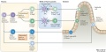
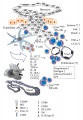
 Intestinal function and microbiota 01.jpg 1,200 × 845; 207 KB
 Intestine histology 002.jpg 800 × 640; 130 KB
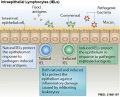
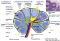
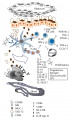
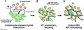
 Intraepithelial lymphocyte differentiation 01.jpg 1,200 × 585; 108 KB
 Intraepithelial lymphocyte differentiation 02.jpg 1,200 × 557; 79 KB
 Intraepithelial lymphocyte differentiation 03.jpg 600 × 484; 68 KB
 Keibel Mall 2 488.jpg 1,200 × 576; 317 KB
 Keibel Mall 2 489.jpg 855 × 1,000; 97 KB
 Keibel Mall 2 490.jpg 614 × 1,000; 198 KB
 Keibel Mall 2 491.jpg 987 × 1,000; 294 KB
 Keibel Mall 2 492.jpg 781 × 1,000; 279 KB
 Keibel Mall 2 493.jpg 680 × 1,000; 89 KB
 Keibel Mall 2 494.jpg 1,200 × 856; 203 KB
 Lymph node - high endothelial venule.jpg 959 × 615; 193 KB
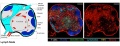
 Lymph node 05.jpg 1,000 × 800; 180 KB
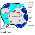
 Lymph node cartoon 01.jpg 800 × 538; 109 KB
 Lymph node cartoon 02.jpg 1,000 × 449; 97 KB
 Lymph node cartoon 03.jpg 1,000 × 673; 297 KB
 Lymph node cartoon.jpg 600 × 450; 66 KB
 Lymph node cells 01.jpg 800 × 617; 142 KB
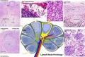
 Lymph node histology 01.jpg 600 × 400; 61 KB
 Lymph node histology 02.jpg 450 × 600; 130 KB
 Lymph node histology 03.jpg 450 × 600; 140 KB
 Lymph node histology 04.jpg 450 × 600; 88 KB
 Lymph node histology 05.jpg 450 × 600; 87 KB
 Lymph node histology 06.jpg 450 × 600; 141 KB
 Lymph node histology01.jpg 800 × 680; 282 KB
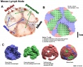 Lymph node model dymamics.jpg 1,000 × 800; 231 KB
 Lymph node structure 01.jpg 1,200 × 463; 167 KB
 Lymph node structure 01.png 1,000 × 820; 395 KB
 Lymph node structure 02.jpg 460 × 463; 54 KB
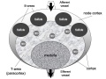
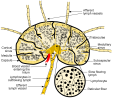
 Lymph node structure.jpg 700 × 418; 52 KB
 Lymph nodes head neck superficial.jpg 419 × 400; 13 KB
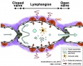
 Lymphatic capillary.jpg 690 × 400; 69 KB
 Lymphatic microvasculature model.jpg 356 × 440; 40 KB
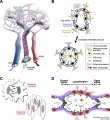 Lymphatic vasculature 01.jpg 1,177 × 1,280; 249 KB
 Lymphatic vasculature 02.jpg 1,280 × 454; 116 KB
 Lymphatic vasculature 03.jpg 713 × 563; 75 KB
 Lymphatic vasculature 04.jpg 560 × 766; 76 KB
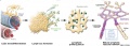
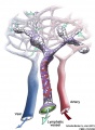
 Lymphatic vessel development.jpg 600 × 410; 92 KB
 Lymphatic vessel formation model.jpg 600 × 692; 179 KB
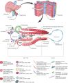
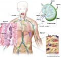
 Lymphatic-system-bone-marrow.jpg 800 × 755; 143 KB
 Lymphatic-system-node.jpg 800 × 755; 139 KB
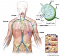
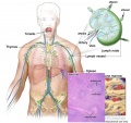
 Lymphatic-system-overview.jpg 800 × 755; 118 KB
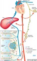
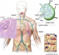
 Lymphatic-system-spleen.jpg 800 × 755; 139 KB
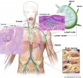
 Lymphatic-system-thymus.jpg 800 × 755; 144 KB
 Lymphatic-system-tonsil-MALT.jpg 800 × 755; 144 KB
 Lymphatic-system-tonsil.jpg 800 × 755; 130 KB
 Lymphocyte rosettes EM01-06.jpg 1,364 × 2,100; 334 KB
 Lymphocyte rosettes EM01.jpg 661 × 665; 58 KB
 Lymphocyte rosettes EM012.jpg 618 × 661; 59 KB
 Lymphocyte rosettes EM02.jpg 661 × 665; 62 KB
 Lymphocyte rosettes EM03.jpg 661 × 665; 51 KB
 Lymphocyte rosettes EM04.jpg 661 × 665; 53 KB
 Lymphocyte rosettes EM05.jpg 661 × 665; 55 KB
 Lymphocyte rosettes EM06.jpg 661 × 665; 66 KB
 Model for immune role in implantation - Mucin absence.jpg 600 × 870; 213 KB
 Model for immune role in implantation.jpg 600 × 1,001; 231 KB
 Mouse adult lymph node 01.mov ; 1.58 MB
- Mouse adult lymph node 02.mov ; 2.3 MB
- Mouse adult lymph node 03.mov ; 1.11 MB
- Mouse adult lymph node 04.mov ; 568 KB
- Mouse adult lymph node 05.mov ; 1.41 MB
- Mouse adult lymph node 06.mov ; 2.17 MB
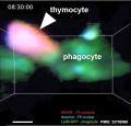 Mouse adult thymus 11.jpg 510 × 490; 34 KB
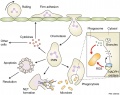 Neutrophil function model 01.jpg 1,203 × 949; 134 KB
 Neutrophil function model 02.jpg 1,220 × 1,243; 166 KB
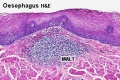 Oesophagus MALT.jpg 500 × 333; 73 KB
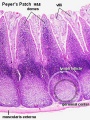 Peyer's patch 01.jpg 450 × 600; 118 KB
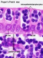 Peyer's patch 02.jpg 450 × 600; 69 KB
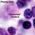 Plasma cell clockface nucleus 01.jpg 400 × 400; 27 KB
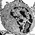 Plasma cell EM06.jpg 595 × 600; 84 KB
 Reticular network formation model.jpg 1,106 × 406; 100 KB
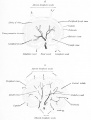
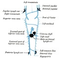
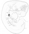
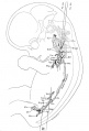
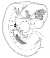
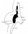
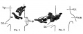 Sabin1909 fig01-02.jpg 636 × 262; 19 KB
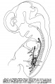
 Sabin1909 fig04.jpg 769 × 902; 97 KB
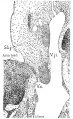
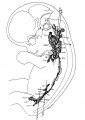
 Sabin1909 fig09.jpg 661 × 800; 34 KB
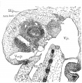
 Sabin1909 fig11.jpg 702 × 1,076; 109 KB
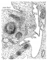
 Sabin1909 fig12.jpg 713 × 1,003; 89 KB
 Sabin1909 fig13.jpg 640 × 547; 114 KB
 Sabin1909 fig16.jpg 512 × 438; 120 KB
 Sabin1909 fig17.jpg 645 × 545; 80 KB
 SH Lecture 180219 Lymphatics.mp3 ; 18.62 MB
 Spleen anatomy.jpg 275 × 285; 19 KB
 Spleen histology 01.jpg 450 × 600; 133 KB
 Spleen histology 02.jpg 455 × 606; 142 KB
 Spleen histology 03.jpg 450 × 600; 83 KB
 Spleen histology 04.jpg 450 × 600; 108 KB
 Spleen histology 05.jpg 450 × 600; 170 KB
 Spleen histology 06.jpg 1,000 × 800; 408 KB
 Spleen histology 07.jpg 1,000 × 800; 251 KB
 Spleen histology 08.jpg 1,000 × 800; 304 KB
 Spleen histology 09.jpg 1,280 × 1,024; 692 KB
 Spleen histology 10.jpg 1,280 × 1,024; 444 KB
 Spleen histology 11.jpg 1,280 × 1,024; 410 KB
 Spleen histology 12.jpg 600 × 450; 93 KB
 Spleen histology 13.jpg 600 × 450; 80 KB
 Stage 22 image 169.jpg 1,000 × 650; 192 KB
 T and B lymphocytes EM09.jpg 1,196 × 627; 137 KB
 T and B lymphocytes EM10.jpg 1,196 × 677; 155 KB
 T2 lymphocyte EM13.jpg 781 × 795; 177 KB
 T2 lymphocyte EM14.jpg 728 × 771; 156 KB
 Thymus histology 01.jpg 1,280 × 1,024; 723 KB
 Tonsil histology 01.jpg 450 × 600; 106 KB
 Tonsil histology 02.jpg 450 × 600; 62 KB


