Category:Guinea Pig: Difference between revisions
From Embryology
(Created page with 'This page lists UNSW Embryology content related to guinea pig development.') |
No edit summary |
||
| Line 1: | Line 1: | ||
This page lists UNSW Embryology content related to guinea pig development. | This page lists UNSW Embryology content related to guinea pig development. | ||
[[Category:Animal Development]] | |||
Revision as of 11:46, 14 July 2012
This page lists UNSW Embryology content related to guinea pig development.
Pages in category 'Guinea Pig'
The following 28 pages are in this category, out of 28 total.
P
- Template:Papanicolaou1933 guinea pig and rat vaginal smear table
- Paper - A contribution to the early development of the heart in mammalia, with special reference to the guinea pig
- Paper - Changes in the vaginal epithelium of the guinea-pig during the oestrous cycle (1922)
- Paper - Growth of the reproductive and endocrine organs of the guinea-pig (1936)
- Paper - Guinea pig development 11 to 21 days
- Paper - Guinea pig development 21 to 35 days
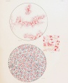
- Paper - Studies on guinea pig oocytes 1
- Paper - The embedding of the embryo guinea-pig in the uterine wall and its nutrition at that stage of development
- Paper - The existence of a typical oestrous cycle in the guinea-pig (1917)
R
- Template:Ref-AdamsHertig1964
- Template:Ref-DeaneslyRowlands1936
- Template:Ref-Emrys-Roberts1910
- Template:Ref-Harman1932
- Template:Ref-Harman1933
- Template:Ref-Huber1918
- Template:Ref-League1928
- Template:Ref-Marion1913
- Template:Ref-Nicol1933
- Template:Ref-Selle1922
- Template:Ref-Silva2016
- Template:Ref-Stockard1917
- Template:Ref-Wilson1928
- Template:Ref-Yoshinaga1921
Media in category 'Guinea Pig'
The following 12 files are in this category, out of 12 total.
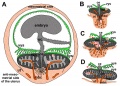 Fetal membrane and placenta cartoon.jpg 600 × 429; 125 KB
 Guineapig icon.jpg 240 × 180; 7 KB
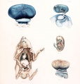
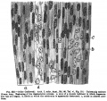 Keibel Mall 332.jpg 851 × 800; 142 KB
 Placenta humans and guinea-pig cartoon.jpg 1,200 × 889; 562 KB
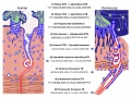
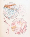
 Placental trophospongium.jpg 567 × 344; 94 KB
 Salvi1898 fig01-15.jpg 2,500 × 1,836; 535 KB
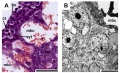
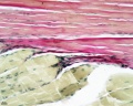 Skeletal muscle histology 004.jpg 1,280 × 1,024; 242 KB
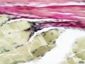 Skeletal muscle histology 444.jpg 934 × 701; 125 KB
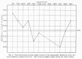 Stockard Papanicolaou1917 figA.jpg 1,382 × 1,000; 143 KB
 Wislocki1920 plate 1.jpg 1,145 × 1,200; 173 KB
 Wislocki1920 plate 2.jpg 988 × 1,200; 245 KB
 Wislocki1920 plate 3.jpg 975 × 1,200; 219 KB


