Category:Genital
From Embryology
This Embryology category shows pages and media related to development of the genital system.
Subcategories
This category has the following 26 subcategories, out of 26 total.
A
C
D
F
M
T
U
V
Pages in category 'Genital'
The following 200 pages are in this category, out of 430 total.
(previous page) (next page)2
- 2009 Lecture 16
- 2009 Lecture 20
- 2010 Lab 8
- 2010 Lecture 19
- 2011 Lab 8
- 2011 Lab 8 - Early Embryo
- 2011 Lab 8 - Fetal
- 2011 Lab 8 - Genital - Quiz
- 2011 Lab 8 - Genital Abnormalities
- 2011 Lab 8 - Late Embryo
- 2011 Lab 8 - Online Assessment
- 2011 Lab 8 - Postnatal
- 2011 Lab 8 - Sex Determination
- Template:2011Lab8
- Template:2016BGDLecture-Genital
- 2016BGDLecture-Genital-Movie
- Template:2016Lecture-Genital
- 2016Lecture-Genital-Movie
A
- Template:AMH
- ANAT2241 Male Reproductive System
- Talk:ANAT2341 Lab 1 - Sex Determination
- ANAT2341 Lab 8 - Early Embryo
- ANAT2341 Lab 8 - Fetal
- ANAT2341 Lab 8 - Genital Abnormalities
- ANAT2341 Lab 8 - Late Embryo
- ANAT2341 Lab 8 - Online Assessment
- ANAT2341 Lab 8 - Postnatal
- ANAT2341 Lab 8 - Sex Determination
- Template:ANAT2341Lab8
- Template:Anorchia
- Template:Anti-Mullerian Hormone
- Template:Anti-Mullerian Hormone Vignette
- ANZACA Meeting 2012 - Embryology
- Atlas of the Development of Man 2 - Urogenital
- Audio
B
- BGD Lecture - Sexual Differentiation
- BGD Practical - Sexual Differentiation Quiz
- BGDB Practical - Sexual Differentiation
- Talk:BGDB Practical - Sexual Differentiation
- BGDB Practical - Sexual Differentiation Interactive
- Template:BGDB SexDiffn
- BGDB Sexual Differentiation - Abnormalities
- Template:BGDB Sexual Differentiation - Abnormalities Interactive
- BGDB Sexual Differentiation - Early Embryo
- Template:BGDB Sexual Differentiation - Early Embryo Interactive
- BGDB Sexual Differentiation - Fetal
- Template:BGDB Sexual Differentiation - Fetal Interactive
- BGDB Sexual Differentiation - Late Embryo
- Template:BGDB Sexual Differentiation - Late Embryo Interactive
- BGDB Sexual Differentiation - Postnatal
- Template:BGDB Sexual Differentiation - Postnatal Interactive
- BGDB Sexual Differentiation - Quiz
- BGDB Sexual Differentiation - Sex Determination
- Template:BGDB Sexual Differentiation - Sex Determination Interactive
- Book - A Laboratory Manual and Text-book of Embryology 8
- Book - A text-book of experimental cytology (1931)
- Book - Brain and behavioural development 8
- Book - Contributions to Embryology Carnegie Institution No.54
- Book - Contributions to Embryology Carnegie Institution No.61
- Book - Educational History of the Genitalia
- Book - Manual of Human Embryology 19
- Book - Manual of Human Embryology 19-2
- Book - Manual of Human Embryology 19-3
- Book - Manual of Human Embryology 19-4
- Book - Quain's Embryology 10
- Book - Sex and internal secretions (1961)
- Book - Text-Book of Embryology 15
- Book - The mammary apparatus of the mammalia (1920)
- Template:Bulbourethral gland
C
- Template:Cloaca
- Cloaca Development
- Template:Cloacal exstrophy
- Template:Cloacal membrane
- Template:Collapsible Table Mullerian Duct Growth
- Template:Collapsible Table Reproductive Gland Growth
- Template:Congenital adrenal hyperplasia
- Template:Corpus albicans
- Template:Corpus luteum
- Corpus Luteum Development
- Template:Cryptorchidism
- Template:Cryptorchidism table
D
E
G
- Template:Genital
- Genital - Female Development
- Genital - Male Development
- Template:Genital abnormalities
- Genital Abnormality - Hypospadias
- Template:Genital cartoons
- Template:Genital Links
- Genital Quiz
- Genital System - Abnormalities
- Talk:Genital System - Abnormalities
- Genital System Development
- Template:Genital terms
- Template:Germinal epithelium
- Template:Gonad
- Gonad Blood Supply Development
- Template:Graafian
- Template:Graafian follicle
- Template:Granulosa lutein cells
- Template:Gray1110 links
- Template:Gubernaculum
H
I
L
M
O
P
- Paper - A contribution to the development of the prostate in man (1909)
- Paper - A morphological study of testicular descent
- Paper - A note on a case of bifid penis (1924)
- Paper - A note on the origin and histogenesis of the mesonephric duct in mammals
- Paper - A previllous human embryo (1946)
- Paper - A report on two cases of hermaphroditism in man (1923)
- Paper - A study of the function of the epididymis 1 (1929)
- Paper - A suggestion as to the cause of the aspermatic condition of the imperfectly descended testis (1922)
- Paper - Adult human ovaries with follicles containing several oocytes (1912)
- Paper - An experimental study of the morphogenesis of intersexuality (1940)
- Paper - Anatomy, pathology and development of the hymen
- Paper - Anomalies of the genito-urinary tract
- Paper - Another case of hermaphroditism in man (1924)
- Paper - Chiefly concerning the genito-mesenteric fold of peritoneum
- Paper - Cysts of the genital ducts Müllerian and Wolffian (1946)
- Paper - Cytology of the human spermatozoon
- Paper - Development and transition of the testis, normal and abnormal 1
- Paper - Development and transition of the testis, normal and abnormal 2
- Paper - Development and transition of the testis, normal and abnormal 3
- Paper - Development and transition of the testis, normal and abnormal 4
- Paper - Development and vascularization of the testis (1906)
- Paper - Development of the egg of the cow up to the stage of blastocyst formation (1946)
- Paper - Development of the human ovary from birth to sexual maturity
- Paper - Development of the mammary gland
- Paper - Development of the mammary gland - Arris and Gale Lecture
- Paper - Development of the Mouse Gonads 1
- Paper - Development of the Mouse Gonads 2
- Paper - Development of the Mouse Gonads 3
- Paper - Development of the Mouse Gonads 4
- Paper - Development of the trigone of the bladder and the termination of the mesonephric ducts (1946)
- Paper - Development of the urogenital system in the Marsupialia 2
- Paper - Development of the uterine glands in man (1920)
- Paper - Development of the vagina in the human fetus
- Paper - Electron microscopy of the sperm tail - results obtained with a new fixative
- Paper - Experimental evidence regarding the role of the anterior pituitary in the development and regulation of the genital system
- Paper - Growth of the reproductive and endocrine organs of the guinea-pig (1936)
- Paper - Has a persistence of the Müllerian ducts any relation to the conditions of cryptorchidism?
- Paper - Hermaphroditism in a mole with male external genitals (1924)
- Paper - Hermaphroditism in man (1920)
- Paper - Histochemical observations on the germ cells of human embryos
- Paper - Histochemical observations on the germ cells of human embryos (1953)
Media in category 'Genital'
The following 200 files are in this category, out of 510 total.
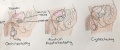
(previous page) (next page)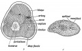 Keith1902 fig111.jpg 1,000 × 620; 145 KB
 Keith1902 fig112.jpg 600 × 541; 77 KB
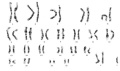 Klinefelter syndrome karyotype.jpg 480 × 284; 12 KB
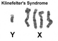 Klinefelter syndrome YXXXX.jpg 244 × 172; 5 KB
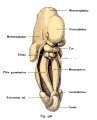 Kollmann426.jpg 500 × 620; 46 KB
 Kollmann445.jpg 776 × 738; 112 KB
 Kollmann446.jpg 723 × 414; 38 KB
 Kollmann447.jpg 564 × 556; 42 KB
 Kollmann448.jpg 643 × 576; 43 KB
 Kollmann449.jpg 613 × 617; 44 KB
 Kollmann450.jpg 757 × 634; 98 KB
 Kollmann451.jpg 796 × 826; 102 KB
 Kollmann452.jpg 687 × 606; 74 KB
 Kollmann453.jpg 664 × 566; 65 KB
 Kollmann454.jpg 729 × 641; 84 KB
 Kollmann455.jpg 746 × 509; 102 KB
 Kollmann458.jpg 1,000 × 520; 120 KB
 Leydig cell PMID13693345 EM02.jpg 1,359 × 957; 341 KB
 Leydig cell PMID13693345 EM03.jpg 1,359 × 957; 325 KB
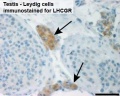 Leydig cells stained for LHCGR1.jpg 404 × 322; 16 KB
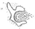 Lockwood1887b fig23.jpg 500 × 423; 41 KB
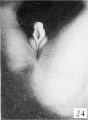
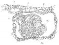 Lockwood1887b fig24.jpg 600 × 463; 82 KB
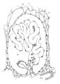 Lockwood1887b fig25.jpg 715 × 1,000; 125 KB
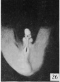
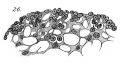 Lockwood1887b fig26.jpg 500 × 266; 38 KB
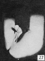
 Lockwood1887b fig27.jpg 500 × 378; 46 KB
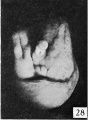
 Lockwood1887b fig28.jpg 600 × 592; 43 KB
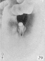
 Lockwood1887b fig29.jpg 800 × 656; 121 KB
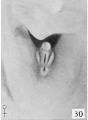
 Lockwood1887b fig30.jpg 800 × 619; 69 KB
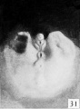
 Lockwood1887b fig31.jpg 800 × 576; 49 KB
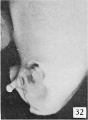
 Lockwood1887b fig32.jpg 500 × 329; 55 KB
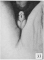
 Lockwood1887b fig33.jpg 600 × 617; 101 KB
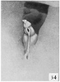
 Lockwood1887b fig34.jpg 408 × 800; 43 KB
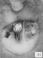
 Lockwood1887b fig35.jpg 800 × 600; 117 KB
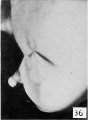
 Lockwood1887b fig36.jpg 600 × 828; 53 KB
 Lockwood1887b fig37.jpg 800 × 739; 102 KB
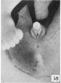
 Lockwood1887b fig38.jpg 800 × 688; 65 KB
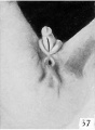
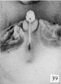
 Lockwood1887b fig39.jpg 800 × 1,102; 93 KB
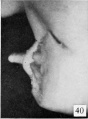
 Lockwood1887b fig40.jpg 774 × 1,000; 92 KB
 Lockwood1887b fig41.jpg 800 × 547; 129 KB
 Lockwood1887b plate02.jpg 2,998 × 2,272; 1.21 MB
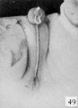
 Lockwood1888a fig47.jpg 1,000 × 712; 279 KB
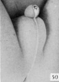
 Lockwood1888a fig50.jpg 959 × 652; 85 KB
 Lockwood1888a plate07.jpg 1,280 × 985; 285 KB
 Lockwood1888b fig49.jpg 800 × 496; 69 KB
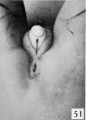
 Lockwood1888b fig51.jpg 800 × 725; 88 KB
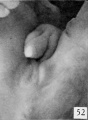
 Lockwood1888b fig52.jpg 800 × 824; 60 KB
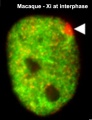 Macaque Xi at interphase 02.jpg 273 × 355; 18 KB
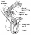 Male - inguinal hernia.jpg 361 × 400; 34 KB
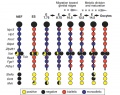
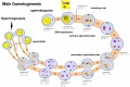 Male gametogenesis.jpg 1,000 × 666; 121 KB
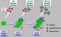
 Male genital molecular signaling 01.jpg 1,232 × 1,032; 94 KB
 Male histology 002.jpg 1,280 × 1,024; 490 KB
 Male histology 003.jpg 1,280 × 1,024; 703 KB
 Male histology 004.jpg 1,280 × 1,024; 540 KB
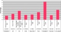
 Male testosterone and AMH level graph.jpg 683 × 360; 31 KB
 Male vas deferens and bladder week6to10.jpg 800 × 918; 86 KB
 Mesonephric duct position week 6-11.jpg 697 × 800; 72 KB
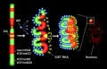 Model for XIST RNA spread from X inactivation center.jpg 1,000 × 642; 104 KB
 Model male androsterone synthesis.jpg 740 × 518; 92 KB
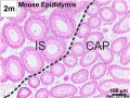
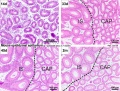 Mouse epididymis development 01.jpg 1,200 × 909; 486 KB
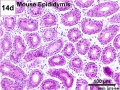 Mouse epididymis development 02.jpg 600 × 451; 138 KB
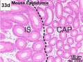 Mouse epididymis development 03.jpg 600 × 451; 119 KB
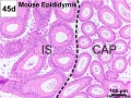 Mouse epididymis development 04.jpg 600 × 451; 120 KB
 Mouse epididymis development 05.jpg 600 × 451; 112 KB
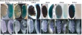
 Mouse external genital development.jpg 800 × 701; 75 KB
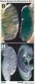
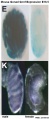
 Mouse fragilis2 expression.jpg 600 × 675; 60 KB
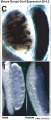
 Mouse genital ridge fragilis expression.jpg 600 × 457; 33 KB
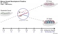 Mouse gonad development timeline.jpg 1,200 × 697; 98 KB
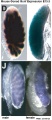
 Mouse gonad Gcnf expression 01.jpg 1,947 × 843; 304 KB
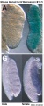 Mouse gonad Gcnf expression E12.5.jpg 331 × 785; 68 KB
 Mouse gonad Gcnf expression E13.5.jpg 332 × 784; 60 KB
 Mouse gonad Gcnf expression E14.5.jpg 334 × 784; 60 KB
 Mouse gonad Gcnf expression E15.5.jpg 338 × 782; 53 KB
 Mouse gonad Gcnf expression E16.5.jpg 325 × 786; 40 KB
 Mouse gonad Gcnf expression E17.5.jpg 328 × 786; 44 KB
 Mouse gonad sex determination 01.jpg 600 × 600; 81 KB
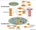
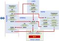
 Mouse sex determination genes 01.jpg 1,280 × 923; 73 KB
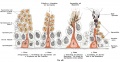
 Mouse- gonadal supporting cell development.jpg 1,000 × 588; 74 KB
 Mouse- spermatozoa NANOG expression.jpg 800 × 512; 111 KB
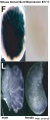
 Mouse- X-linked gene expression in primordial germ cells.jpg 800 × 632; 90 KB
 Mouse-model ovarian cord formation.jpg 600 × 368; 48 KB
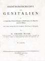 Muller 1850 titlepage.jpg 743 × 1,000; 64 KB
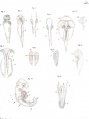 Muller1850 Plate1.jpg 1,490 × 2,000; 265 KB
 Nelsen1953 fig002.jpg 1,200 × 1,037; 249 KB
 Nelsen1953 fig004.jpg 1,200 × 960; 200 KB
 Nelsen1953 fig005.jpg 1,200 × 709; 102 KB
 Nelsen1953 fig006.jpg 1,200 × 732; 224 KB
 Nelsen1953 fig007.jpg 1,200 × 1,311; 343 KB
 Nelsen1953 fig022.jpg 1,200 × 839; 207 KB
 Nelsen1953 fig030.jpg 1,200 × 995; 414 KB
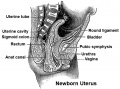 Newborn uterus.jpg 540 × 400; 72 KB
 Newborn- cryptorchidism normal birthweight.jpg 800 × 428; 47 KB
 OHVIRA syndrome 02.jpg 700 × 669; 72 KB
 OHVIRA syndrome 03.jpg 700 × 669; 65 KB
 Orchidometer.jpg 361 × 225; 14 KB
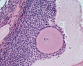
 Ovary histology 001.jpg 1,280 × 1,024; 360 KB
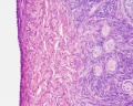
 Ovary histology 002.jpg 1,280 × 1,024; 270 KB
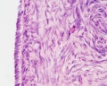
 Ovary histology 003.jpg 1,280 × 1,024; 337 KB
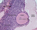
 Ovary histology 004.jpg 1,280 × 1,024; 401 KB
 Ovary histology 005.jpg 1,280 × 1,024; 354 KB
 Ovary histology 006.jpg 1,280 × 1,024; 424 KB
 Ovary histology 007.jpg 1,280 × 1,024; 336 KB
 Ovary histology 008.jpg 1,280 × 1,024; 264 KB
 Ovary histology 061.jpg 1,280 × 1,024; 438 KB
 Ovary histology 061a.jpg 800 × 640; 200 KB
 Ovary histology 061c.jpg 400 × 320; 56 KB
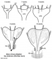
 Paramesonephric duct.jpg 423 × 478; 40 KB
 Paramesonephric ducts.jpg 371 × 400; 43 KB
 Perineal fistula.jpg 800 × 596; 82 KB
 Persistent cloaca perineum.jpg 600 × 800; 57 KB
 Persistent Mullerian duct syndrome 01.jpg 755 × 1,000; 53 KB
 Primordial germ cell 001.mov ; 680 KB
- Primordial germ cell 002.mov ; 3.7 MB
- Primordial germ cell 003.mov ; 695 KB
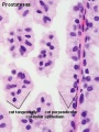
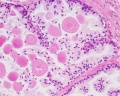
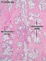 Prostate histology 01.jpg 300 × 400; 72 KB
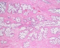
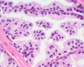
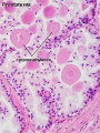 Prostate histology 02.jpg 300 × 400; 57 KB
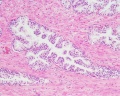
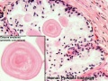
 Prostate histology 03.jpg 300 × 400; 41 KB
 Prostate histology 04.jpg 1,280 × 1,024; 569 KB
 Prostate histology 05.jpg 1,280 × 1,024; 418 KB
 Prostate histology 06.jpg 1,280 × 1,024; 348 KB
 Prostate histology 07.jpg 1,280 × 1,024; 328 KB
 Prostate histology 08.jpg 1,280 × 1,024; 252 KB
 Prostate histology 09.jpg 1,019 × 764; 199 KB
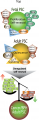 Prostate stem cell cartoon.png 907 × 2,789; 1.09 MB
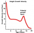 Puberty growth.jpg 528 × 511; 36 KB
 Rat blood–testis barrier 01.jpg 1,002 × 1,599; 221 KB
 Rat blood–testis barrier 02.jpg 1,002 × 853; 125 KB
 Regnier de Graaf.jpg 164 × 202; 6 KB
 Reproductive surgeries in Males.jpeg 1,624 × 676; 354 KB
 Rugh 152.jpg 800 × 669; 122 KB
 Rugh 155.jpg 720 × 1,000; 266 KB
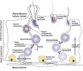
 Seminiferous-tubule-HEx40.jpg 400 × 500; 59 KB
 Septate uterus ultrasound 01.jpg 1,280 × 525; 95 KB
 Simkins1928 plate01.jpg 1,574 × 2,003; 237 KB
 Simkins1928 plate02.jpg 1,464 × 2,126; 223 KB
 Simkins1928 plate03.jpg 1,551 × 2,086; 293 KB
 Simkins1928 plate04.jpg 1,543 × 2,026; 219 KB
 Simkins1928 plate05.jpg 1,281 × 2,111; 154 KB
 Simkins1928 plate06.jpg 1,587 × 2,088; 277 KB
 Simkins1928 plate07.jpg 1,540 × 2,096; 239 KB
 Simkins1928 plate08.jpg 1,558 × 1,797; 220 KB
 Simkins1928 plate09.jpg 1,548 × 2,096; 263 KB
 Simkins1928 plate10.jpg 1,565 × 1,386; 140 KB
 Smooth muscle histology 007.jpg 1,280 × 1,024; 260 KB
 Smooth muscle histology 008.jpg 1,280 × 1,024; 629 KB
 Smooth muscle histology 009.jpg 1,280 × 1,024; 307 KB
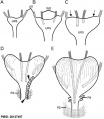
 Spaulding-fig01.jpg 934 × 743; 101 KB
 Spaulding-fig02.jpg 646 × 748; 67 KB
 Spaulding-fig03.jpg 646 × 748; 83 KB
 Spaulding-fig04.jpg 933 × 750; 110 KB
 Spaulding-fig05.jpg 778 × 744; 96 KB
 Spaulding-fig06.jpg 778 × 744; 85 KB
 Spaulding-fig07.jpg 445 × 607; 35 KB
 Spaulding-fig08.jpg 445 × 607; 37 KB
 Spaulding-fig09.jpg 445 × 607; 37 KB
 Spaulding-fig10.jpg 445 × 607; 34 KB
 Spaulding-fig11.jpg 445 × 607; 36 KB
 Spaulding-fig12.jpg 445 × 607; 25 KB
 Spaulding-fig13.jpg 445 × 607; 36 KB
 Spaulding-fig14.jpg 445 × 607; 35 KB
 Spaulding-fig15.jpg 445 × 607; 38 KB
 Spaulding-fig16.jpg 445 × 607; 38 KB
 Spaulding-fig17.jpg 445 × 607; 36 KB
 Spaulding-fig18.jpg 445 × 607; 36 KB
 Spaulding-fig19.jpg 445 × 607; 36 KB
 Spaulding-fig20.jpg 445 × 607; 37 KB
 Spaulding-fig21.jpg 445 × 607; 33 KB
 Spaulding-fig22.jpg 445 × 607; 32 KB
 Spaulding-fig23.jpg 443 × 604; 37 KB
 Spaulding-fig24.jpg 443 × 604; 36 KB
 Spaulding-fig25.jpg 443 × 604; 33 KB
 Spaulding-fig26.jpg 443 × 604; 31 KB
 Spaulding-fig27.jpg 443 × 604; 32 KB
 Spaulding-fig28.jpg 443 × 604; 37 KB
 Spaulding-fig29.jpg 443 × 604; 29 KB
 Spaulding-fig30.jpg 443 × 604; 27 KB
 Spaulding-fig31.jpg 443 × 604; 31 KB
 Spaulding-fig32.jpg 443 × 604; 31 KB
 Spaulding-fig33.jpg 443 × 604; 29 KB
 Spaulding-fig34.jpg 443 × 604; 30 KB
 Spaulding-fig35.jpg 443 × 604; 39 KB
 Spaulding-fig36.jpg 443 × 604; 28 KB
 Spaulding-fig37.jpg 443 × 604; 27 KB
 Spaulding-fig38.jpg 443 × 604; 34 KB
 Spaulding-fig39.jpg 436 × 603; 31 KB
 Spaulding-fig40.jpg 445 × 604; 31 KB
 Spaulding-fig41.jpg 446 × 605; 27 KB
 Spaulding-fig42.jpg 430 × 599; 26 KB
 Spaulding-fig43.jpg 436 × 603; 29 KB
 Spaulding-fig44.jpg 445 × 604; 27 KB
 Spaulding-fig45.jpg 440 × 599; 31 KB
 Spaulding-fig46.jpg 430 × 599; 26 KB
 Spaulding-fig47.jpg 436 × 603; 32 KB
 Spaulding-fig48.jpg 445 × 604; 33 KB
 Spaulding-fig49.jpg 436 × 602; 26 KB
 Spaulding-fig50.jpg 430 × 599; 24 KB
 Spaulding-fig51.jpg 429 × 599; 32 KB
 Spaulding-fig52.jpg 439 × 601; 33 KB
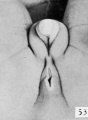 Spaulding-fig53.jpg 436 × 596; 28 KB
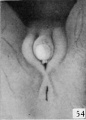 Spaulding-fig54.jpg 430 × 599; 27 KB
 Spaulding-plate01.jpg 1,266 × 2,000; 392 KB


