Category:Genital: Difference between revisions
From Embryology
mNo edit summary |
mNo edit summary |
||
| Line 1: | Line 1: | ||
This {{Embryology}} category shows pages and media related to development of the genital system. | This {{Embryology}} category shows pages and media related to development of the genital system. Note that links below beginning with "Paper -" are generally historic research articles. | ||
{{Genital Links}} | {{Genital Links}} | ||
Latest revision as of 13:46, 14 February 2017
This Embryology category shows pages and media related to development of the genital system. Note that links below beginning with "Paper -" are generally historic research articles.
Subcategories
This category has the following 26 subcategories, out of 26 total.
A
C
D
F
M
T
U
V
Pages in category 'Genital'
The following 30 pages are in this category, out of 430 total.
(previous page) (next page)T
- Template:Table Reproductive Gland Growth
- Template:Tanner stages
- Template:Tanner stages collapse table
- Template:Testis
- Testis Descent Movie
- Testis Development
- Testis Development Movie
- Template:Testis Histology
- Template:Testosterone
- Template:Theca lutein cells
- Trigone Development Movie
- Template:Tunica albuginea
U
Media in category 'Genital'
The following 200 files are in this category, out of 510 total.
(previous page) (next page) Allen1904 plate7.jpg 1,280 × 1,837; 704 KB
 Alternate androgen synthesis pathway.jpg 750 × 425; 99 KB
 Anogenital distance from birth to 2 years.jpg 565 × 545; 34 KB
 Australian abnormalities 81-92 urogenital.jpg 600 × 429; 54 KB
 Azoospermia.jpg 768 × 554; 77 KB
 Bacterial vaginosis.png 1,000 × 755; 134 KB
 Bailey307.jpg 832 × 833; 128 KB
 Bailey308.jpg 791 × 848; 88 KB
 Bailey309.jpg 594 × 592; 58 KB
 Bailey310.jpg 688 × 365; 41 KB
 Bailey327.jpg 872 × 567; 89 KB
 Bailey328.jpg 704 × 795; 85 KB
 Bailey329.jpg 838 × 381; 70 KB
 Bailey330.jpg 680 × 539; 109 KB
 Bailey331.jpg 890 × 782; 118 KB
 Bailey332.jpg 637 × 356; 53 KB
 Bailey333.jpg 830 × 445; 74 KB
 Bailey334.jpg 857 × 558; 77 KB
 Bailey335.jpg 590 × 468; 55 KB
 Bailey336.jpg 912 × 597; 145 KB
 Bailey337.jpg 790 × 573; 74 KB
 Bailey338.jpg 940 × 473; 87 KB
 Bailey339.jpg 802 × 602; 111 KB
 Bailey341.jpg 832 × 675; 69 KB
 Bailey342.jpg 777 × 561; 55 KB
 Bailey343-344.jpg 1,185 × 630; 91 KB
 Bailey345-346.jpg 1,193 × 491; 79 KB
 Bailey347-348.jpg 1,198 × 526; 83 KB
 Baileytable06.jpg 800 × 258; 27 KB
 Baileytable07.jpg 960 × 833; 152 KB
 BGDB2011-Embryo Lab 010611-01.mp3 ; 3.46 MB
- BGDB2011-Embryo Lab 010611-02.mp3 ; 2.99 MB
- BGDB2011-Embryo Lab 010611-03.mp3 ; 2.66 MB
- BGDB2011-Embryo Lab 010611-04.mp3 ; 2.94 MB
- BGDB2011-Embryo Lab 010611-05.mp3 ; 5.17 MB
 Braune 1877 plate 29B.jpg 868 × 1,200; 322 KB
 Braune 1877 plate 30.jpg 857 × 1,200; 295 KB
 Braune 1877 plate 31.jpg 870 × 1,200; 299 KB
 Bulmer1957 plate01.jpg 1,280 × 1,808; 353 KB
 Cat embryo ovary.jpg 505 × 492; 47 KB
 Caudal duplication syndrome.jpg 700 × 599; 47 KB
 Chicken primordial germ cell migration model.jpg 750 × 447; 69 KB
 Corner1920 fig01.jpg 1,000 × 606; 159 KB
 Corner1920 fig02-05.jpg 1,000 × 671; 216 KB
 Corner1920 Plate 1.jpg 756 × 1,000; 222 KB
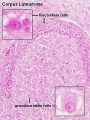 Corpus luteum lutein cells.jpg 450 × 600; 104 KB
 Corpus luteum.jpg 450 × 600; 94 KB
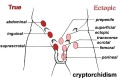 Cryptorchidism.jpg 600 × 390; 35 KB
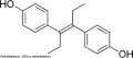 Diethylstilbestrol.jpg 600 × 263; 16 KB
 Dog- spermatozoa NANOG expression.jpg 800 × 691; 109 KB
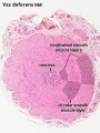 Ductus deferens 01.jpg 400 × 533; 76 KB
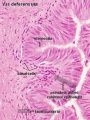 Ductus deferens 02.jpg 400 × 533; 80 KB
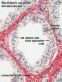
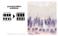 Epididymis histology 01.jpg 600 × 375; 20 KB
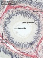
 Epididymis histology 02.jpg 400 × 534; 71 KB
 Epididymis histology 03.jpg 400 × 533; 68 KB
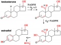 Estradiol synthesis.jpg 600 × 441; 40 KB
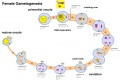 Female gametogenesis.jpg 1,000 × 666; 94 KB
 Female genital and ureter abnormality 01.jpg 766 × 732; 86 KB
 Female genital and ureter abnormality 02.jpg 766 × 733; 78 KB
 Female genital and ureter abnormality 03.jpg 766 × 762; 79 KB
 Female genital tract chlamydia trachomatis infection 01.jpg 804 × 500; 78 KB
 Female reproductive tract Wnt4.jpg 1,000 × 563; 79 KB
 Female- OHVIRA syndrome 01.jpg 340 × 1,000; 76 KB
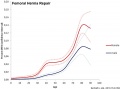 Femoral hernia repair.jpg 800 × 596; 52 KB
 Fetal 10wk urogenital 1.jpg 800 × 600; 109 KB
 Fetal 10wk urogenital 2.jpg 800 × 600; 110 KB
 Fetal 10wk urogenital 3.jpg 800 × 600; 107 KB
 Fetal 10wk urogenital 4.jpg 800 × 600; 105 KB
 Fetal adrenal ectopic germ cells 01.jpg 1,092 × 1,280; 358 KB
 Fetal adrenal ectopic germ cells 02.jpg 1,086 × 446; 124 KB
 Fetal adrenal ectopic germ cells 03.jpg 899 × 700; 159 KB
 Fetal adrenal ectopic germ cells 04.jpg 899 × 700; 147 KB
 Fetal ovary meiosis 01.jpg 1,280 × 410; 132 KB
 Fetal ovary meiosis 02.jpg 496 × 600; 77 KB
 Fetal ovary meiosis 03.jpg 652 × 400; 64 KB
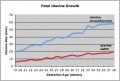 Fetal uterus growth.jpg 438 × 296; 28 KB
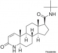 Finasteride.jpg 500 × 463; 18 KB
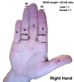 Finger length ratio - 2D4D.jpg 428 × 480; 26 KB
 Fleming1927-fig01.jpg 953 × 1,000; 167 KB
 Fleming1927-fig02.jpg 1,000 × 685; 53 KB
 Fleming1927-fig03.jpg 1,200 × 1,908; 602 KB
 Fleming1927-fig03a.jpg 1,179 × 493; 150 KB
 Fleming1927-fig03b.jpg 1,179 × 577; 171 KB
 Fleming1927-fig03c.jpg 1,179 × 800; 266 KB
 Fleming1927-fig04.jpg 800 × 899; 103 KB
 Fragile X protein cartoon.jpg 1,000 × 615; 107 KB
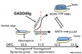 Gadd45g and sex determination model.jpg 909 × 600; 62 KB
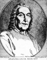 Giovanni Battista Morgagni.jpg 778 × 1,000; 183 KB
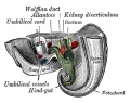
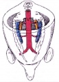 Gonad blood 01 icon.jpg 296 × 413; 28 KB
 Gray0594.jpg 600 × 432; 83 KB
 Gray0619.jpg 800 × 741; 180 KB
 Gray0847.jpg 559 × 900; 155 KB
 Gray0849.jpg 800 × 885; 258 KB
 Gray0982a.jpg 427 × 393; 18 KB
 Gray0982b.jpg 427 × 393; 20 KB
 Gray0986.jpg 565 × 606; 56 KB
 Gray0991.jpg 469 × 400; 33 KB
 Gray0992.jpg 600 × 611; 73 KB
 Gray1108.jpg 590 × 400; 73 KB
 Gray1109.jpg 464 × 487; 56 KB
 Gray1110.jpg 414 × 1,200; 86 KB
 Gray1110a.jpg 578 × 600; 40 KB
 Gray1110b.jpg 578 × 600; 44 KB
 Gray1110c.jpg 578 × 600; 49 KB
 Gray1111.jpg 523 × 600; 66 KB
 Gray1112.jpg 550 × 548; 51 KB
 Gray1113.jpg 600 × 385; 68 KB
 Gray1114.jpg 450 × 471; 47 KB
 Gray1115.jpg 600 × 474; 73 KB
 Gray1116.jpg 600 × 433; 88 KB
 Gray1117.jpg 581 × 510; 75 KB
 Gray1118.jpg 600 × 403; 45 KB
 Gray1119.jpg 700 × 807; 115 KB
 Gray1137.jpg 600 × 502; 64 KB
 Gray1138.jpg 656 × 472; 57 KB
 Gray1139.jpg 600 × 524; 73 KB
 Gray1152.jpg 597 × 600; 100 KB
 Gray1153.jpg 725 × 600; 100 KB
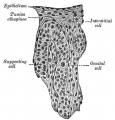
 Historic-ovary.jpg 385 × 283; 34 KB
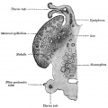
 Historic-testis.jpg 509 × 800; 67 KB
 Human anogenital distance.jpg 570 × 499; 23 KB
 Human Embryology Manual 2 19 - Urogenital fold table.jpg 1,280 × 816; 166 KB
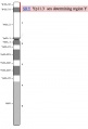
 Human idiogram-chromosome X.jpg 31 × 293; 3 KB
 Human infant ovary follicle 01.jpg 800 × 800; 107 KB
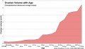
 Human male anogenital distance graph.jpg 600 × 429; 28 KB
 Human ovary postnatal growth.jpg 800 × 467; 40 KB
 Human X chromosome.jpg 400 × 450; 25 KB
 Human Y chromosome 01.jpg 937 × 549; 167 KB
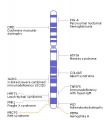
 Human Y chromosome SRY region.jpg 351 × 517; 17 KB
 Human- fetal week 10 lower body A.jpg 600 × 450; 96 KB
 Human- fetal week 10 lower body B.jpg 600 × 450; 93 KB
 Human- fetal week 10 lower body C.jpg 600 × 450; 94 KB
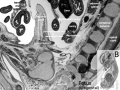
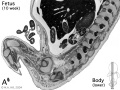
 Human- fetal week 10 urogenital A.jpg 600 × 450; 109 KB
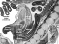
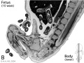
 Human- fetal week 10 urogenital B.jpg 600 × 450; 109 KB
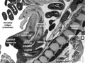
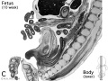
 Human- fetal week 10 urogenital C.jpg 600 × 450; 105 KB
 Human- fetal week 10 urogenital D.jpg 600 × 450; 101 KB
 Human- genital development critical periods.jpg 1,000 × 494; 78 KB
 Human- spermatozoa NANOG expression 01.jpg 798 × 797; 79 KB
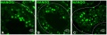 Human- spermatozoa NANOG expression.jpg 1,000 × 333; 77 KB
 Hydrocolpos.jpg 375 × 361; 23 KB
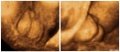 Hypospadia 3D ultrasound 01.jpg 1,150 × 497; 87 KB
 Hypospadia classifications.jpg 500 × 410; 47 KB
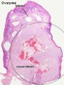
 Infant ovary.jpg 943 × 571; 108 KB
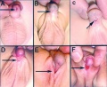
 Inguinal hernia repair 2.jpg 800 × 718; 53 KB
 Inguinal hernia repair.jpg 800 × 597; 46 KB
 Keibel Mall 2 552.jpg 1,280 × 948; 257 KB
 Keibel Mall 2 553.jpg 1,280 × 1,376; 496 KB
 Keibel Mall 2 554a.jpg 1,280 × 933; 221 KB
 Keibel Mall 2 554b.jpg 1,280 × 798; 153 KB
 Keibel Mall 2 554c.jpg 1,280 × 872; 188 KB
 Keibel Mall 2 556.jpg 1,280 × 1,473; 312 KB
 Keibel Mall 2 557.jpg 1,000 × 665; 167 KB
 Keibel Mall 2 558.jpg 1,280 × 792; 132 KB
 Keibel Mall 2 626.jpg 850 × 679; 143 KB
 Keibel Mall 2 627.jpg 1,000 × 1,252; 169 KB
 Keibel Mall 2 628.jpg 1,000 × 512; 65 KB
 Keibel Mall 2 632.jpg 1,000 × 684; 100 KB
 Keibel Mall 2 635.jpg 1,000 × 1,073; 223 KB
 Keibel Mall 2 636.jpg 1,200 × 738; 119 KB
 Keibel Mall 2 641.jpg 1,041 × 518; 62 KB
 Keibel Mall 2 642.jpg 1,200 × 644; 95 KB
 Keibel Mall 2 643.jpg 975 × 1,000; 95 KB
 Keibel Mall 2 645.jpg 957 × 1,000; 98 KB
 Keibel Mall 2 646.jpg 1,280 × 1,307; 181 KB
 Keibel Mall 2 652.jpg 1,200 × 1,036; 130 KB
 Keibel Mall 2 656.jpg 1,200 × 746; 152 KB
 Keibel Mall 2 658a.jpg 1,127 × 1,200; 103 KB
 Keibel Mall 2 658b.jpg 895 × 1,200; 98 KB
 Keibel Mall 2 658c.jpg 1,000 × 1,019; 90 KB
 Keith1902 fig079.jpg 742 × 800; 78 KB
 Keith1902 fig080.jpg 651 × 700; 79 KB
 Keith1902 fig081.jpg 818 × 800; 113 KB
 Keith1902 fig082.jpg 924 × 800; 98 KB
 Keith1902 fig083.jpg 782 × 700; 79 KB
 Keith1902 fig084.jpg 732 × 800; 88 KB
 Keith1902 fig085.jpg 800 × 590; 78 KB
 Keith1902 fig086.jpg 842 × 700; 84 KB
 Keith1902 fig087.jpg 800 × 613; 90 KB
 Keith1902 fig088.jpg 788 × 1,000; 92 KB
 Keith1902 fig089.jpg 964 × 800; 92 KB
 Keith1902 fig090.jpg 700 × 450; 58 KB
 Keith1902 fig091.jpg 700 × 423; 48 KB
 Keith1902 fig093.jpg 700 × 574; 59 KB
 Keith1902 fig094.jpg 800 × 646; 102 KB
 Keith1902 fig095.jpg 660 × 1,000; 125 KB
 Keith1902 fig096.jpg 700 × 535; 69 KB
 Keith1902 fig097.jpg 680 × 592; 80 KB
 Keith1902 fig098.jpg 560 × 460; 40 KB
 Keith1902 fig099.jpg 784 × 800; 105 KB
 Keith1902 fig100.jpg 1,000 × 569; 81 KB
 Keith1902 fig101.jpg 670 × 545; 69 KB
 Keith1902 fig102.jpg 800 × 605; 68 KB
 Keith1902 fig103.jpg 1,000 × 723; 139 KB
 Keith1902 fig104.jpg 800 × 601; 77 KB
 Keith1902 fig105.jpg 632 × 700; 66 KB
 Keith1902 fig108.jpg 780 × 585; 72 KB
 Keith1902 fig109.jpg 859 × 800; 104 KB
 Keith1902 fig110.jpg 800 × 638; 102 KB


