Brain Awareness Week 2012: Difference between revisions
| Line 30: | Line 30: | ||
| [[File:Brain_fissure_development_03.jpg|90px|link=Quicktime Movie - Neural Sylvian Fissure]] | | [[File:Brain_fissure_development_03.jpg|90px|link=Quicktime Movie - Neural Sylvian Fissure]] | ||
| [[File:Adult human brain movie icon.jpg|120px|link=Quicktime_Movie_-_Adult_Brain]] | | [[File:Adult human brain movie icon.jpg|120px|link=Quicktime_Movie_-_Adult_Brain]] | ||
|-bgcolor=" | |-bgcolor="a3bfb1" | ||
| Week 3 | | Week 3 | ||
| Week 4 to 5 | | Week 4 to 5 | ||
| Line 38: | Line 38: | ||
| Week 13 to 21 | | Week 13 to 21 | ||
| Adult Human | | Adult Human | ||
|- | |-bgcolor="F5FFFA" | ||
| [[Quicktime Development Animation - Neural Plate|Neural Plate]] | | [[Quicktime Development Animation - Neural Plate|Neural Plate]] | ||
| [[Quicktime Development Animation - Neural Tube|Neural Tube]] | | [[Quicktime Development Animation - Neural Tube|Neural Tube]] | ||
| [[Quicktime Movie_-_Central_Nervous_System_3D_stage_13|Simple Tube]] | | [[Quicktime Movie_-_Central_Nervous_System_3D_stage_13|Simple Tube]] | ||
| [[Quicktime_Movie_-_Carnegie_Stage_17_Neural| | | [[Quicktime_Movie_-_Carnegie_Stage_17_Neural|Folded Tube]] | ||
| [[Quicktime Movie_-_Central_Nervous_System_3D_stage_22|Central Nervous]] | | [[Quicktime Movie_-_Central_Nervous_System_3D_stage_22|Central Nervous]] | ||
| [[Quicktime Movie - Neural Sylvian Fissure|Fetal Brain]] | | [[Quicktime Movie - Neural Sylvian Fissure|Fetal Brain]] | ||
Revision as of 01:11, 10 March 2012
Welcome to Brain Development
| width=320px|height=260px|controller=false|autoplay=true</qt> | In today's demonstration we will be looking at how the brain develops from a simple tube into the complex folded structure that you will be seeing (and using) today.
This page has been prepared as a simplified introduction to human neural development. |
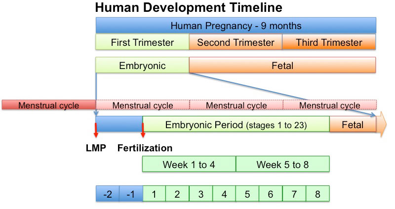
Here is Human Development
This graph shows how we divide human development into different times. Key events occur in the first trimester (embryonic), though the neural system continues to develop through the second and third trimester (fetal) and even after birth (postnatal). This is why it one reason why it is so susceptible to damage.
Here is how the human nervous system grows

|

|
|||||
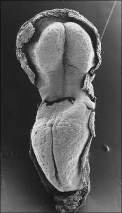
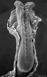
| Week 3 | Week 4 to 5 | Week 5 | Week 6 | Week 8 | Week 13 to 21 | Adult Human |
| Neural Plate | Neural Tube | Simple Tube | Folded Tube | Central Nervous | Fetal Brain | Brain Slices |
Here is a developing mouse nervous system
| width=336px|height=415px|controller=true|autoplay=false</qt> |
This movie shows a 11.5 days old mouse brain. (Mouse development takes 21 days and is a model used in research)
Red - brain
|
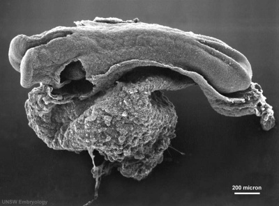
It begins as a Plate
That folds to a Tube
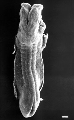
The tube then Closes at each End
These images show the neural tube closing leaving an opening (neuropore) at each end.
|

|
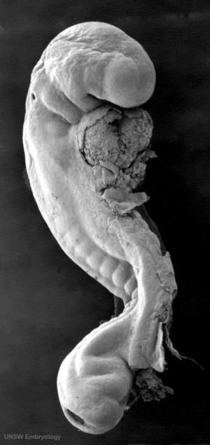
The brain end of the tube forms 3 Vesicles
| width=516px|height=540px|controller=true|loop=true|autoplay=true</qt>
|
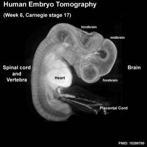
Week 6 - the brain and spinal cord of the human embryo. Also visible are the heart (bright white) and placental cord containing placental blood vessels. |
Brain
At the brain end - the tube expands to form three vesicle (sac or bubble) regions. These will form different parts of the brain and brain stem.
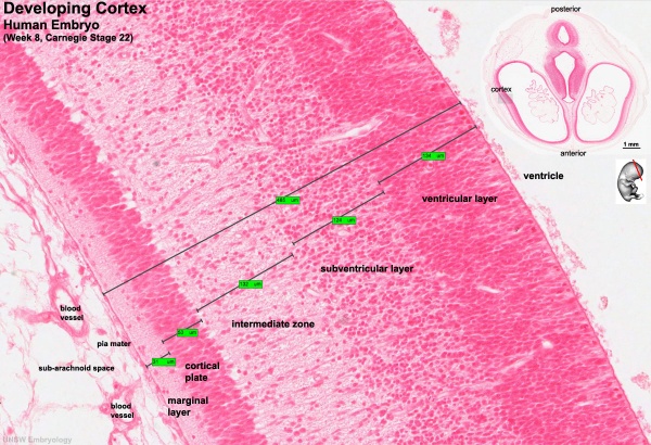
Week 8 wall of the neural tube at the brain end, the smaller images (top right) show the section level from the embryo. The thin layer outer called cortical plate will eventually form the adult brain cortex. The other underlying layers are part of the development process and will continue to supply cells to the cortex through fetal period, these layers will eventually be mainly lost. The ventricle is the fluid-filled space within the brain.
Spinal Cord
At the spinal cord end - the tube stays narrow. This region begins to put out motor nerves to innervate muscle and sensory nerves grow towards the developing spinal cord.
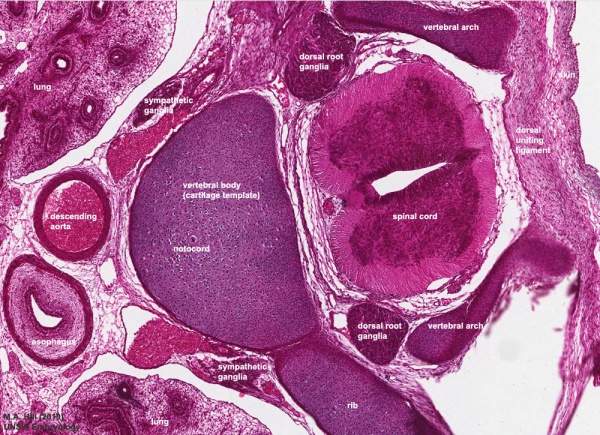
Week 8 wall of the neural tube at the spinal cord end, lying behind the vertebral body. The dark central region is where the neurons (cell bodies, grey matter) are located, the pale outer region is where nerve fibres run (axons, white matter). The dorsal root ganglia lie outside the spinal cord and contain the cell bodies of the sensory neurons.
Fetal brain Grows
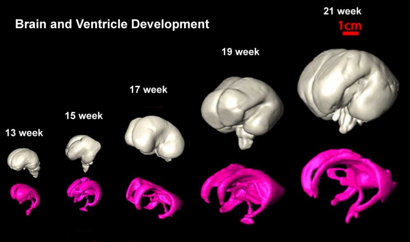
This shows the growth of the brain and the fluid-filled space within the brain (the red bar is 1 cm).
- The brain goes from having a smooth surface to begin to fold or "wrinkle" as the surface area grows.
- The fluid space is filled with cerebrospinal fluid or CSF.
Newborn brain Grows
The brain has not finished growing at birth.
Much of the growth in size after birth is due to "white matter" development, the support cells of the brain, spinal cord and nerves.
