BGDB Sexual Differentiation - Late Embryo
Week 8
| <html5media height="500" width="505">File:Stage22_URG3d.mp4</html5media>
Click Here to play on mobile device Week 8, Carnegie stage 22, male. Page |
Begin by observing the internal structure of the embryo at the end of week 8.
|

|

|
| G5 urogenital | urogenital |
Embryo (week 8, Stage 22) Renal

E6: R,L adrenal glands under diaphragm.
E7: Large adrenal glands. Inferior vena cava. Thoracic aorta. |

|

|

|
| F1: Adrenal glands. R. Kidney. Autonomic ganglia (partly the adrenal medulla precursors). | F2: Kidneys (note retroperitoneal location). Cortex. Medulla. L. Adrenal gland. Superior mesenteric artery. Inferior vena cava. | F3: R testis (note its location relative to the R adrenal). L adrenal. R renal hilus. large channels are branches of ureteric tree. | |

|

|

|

|
| F4: R kidney and R ureter. Inferior vena cava. L. kidney, L renal hilus and L ureter. R testis with R mesonephric duct (precursor of vas deferens). L testis. Umbilical arteries passing into umbilical cord allantois between them. | F5: Kidneys. Ureters. Note umbilical arteries and allantois. Also note how R testis and mesonephric structures are attached to parietal peritoneum by a mesogonad. | F6: Kidneys. Ureters. Note umbilical arteries and allantois. Also note how R testis and mesonephric structures are attached to parietal peritoneum by a mesogonad. | F7: In F7, (dorsal to R testis and liver) note with the distinct lumen of the mesonephric duct, almost solid column of paramesonephric cells and remnants of mesonephric tubules. "mesogonad". Ureters. Bladder with submucosa and detrusor muscle. Umbilical arteries. Division of aorta. |

|

|

|

|
| G1: Ureters, Bladder. Umbilical arteries. Testis with remains of mesonephros (dorsal), mesonephric duct and paramesonephric cells. Sigmoid colon and mesocolon. | G2: Ureters being displaced ventrally, crossing common iliac arteries. Sigmoid colon. Bladder. Mesonephric ducts (lateral) and paramesonephric ducts (smaller, medial) located dorsal to bladder. | G3: Ureters (cut twice): descending dorsal to bladder and ascending ventrally to enter the bladder at trigone, through the submucosa). Fusion of paramesonephric ducts. Paired mesonephric ducts. Umbilical arteries looping off common iliac arteries. Pubic symphysis. Colon. | G4: Most caudal part of loop of ureters. Urethra emerging from bladder. Mesonephric ducts. Rectocolic junction. |

|

|

|
|
| G5: Urethra (in region of future prostate gland - note crescentic shape). Rectum. Rectovesical pouch. Between G4 and G5, each mesonephric duct (vas deferens) has joined the prostatic urethra (caudal to the ureters), thereby increasing the caliber of the latter. | G6: Penile urethra, emerging inferiorly to the glans penis. Scrotal swellings (appear before testis descends). | G7: Penile urethra, emerging inferiorly to the glans penis. Scrotal swellings (appear before testis descends). | Note F7 MS term: "inebriated Puffin" (dorsal to R testis and liver) lumen of the mesonephric duct (eye), almost solid column of paramesonephric cells (beak) and remnants of mesonephric tubules (body). |

Urinary System Development
Development of the Urinary Bladder
| <mediaplayer width='360' height='500' image="http://php.med.unsw.edu.au/embryology/images/f/fe/Urogenital_sinus_001_icon.jpg">File:Urogenital_sinus_001.mp4</mediaplayer> | <mediaplayer width='420' height='415' image="http://php.med.unsw.edu.au/embryology/images/8/88/Urogenital_septum_001_icon.jpg">File:Urogenital_septum_001.mp4</mediaplayer> | Division of the Cloaca
|


Development of the Urethra
Further development of the urinary system outflow tract, the urethra, varies depending on the sex of the embryo.
- Males - the pelvic urethra forms the membranous urethra, the prostatic urethra and penile urethra. (The sex of the above animation and sections is male)
- Females - the pelvic urethra forms the membranous urethra and the vestibule of the vagina.
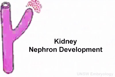
Development of the Kidney
| <mediaplayer width='400' height='280' image="http://php.med.unsw.edu.au/embryology/images/5/59/Renal_001_icon.jpg">File:Nephron_development.mp4</mediaplayer> |
|
| <mediaplayer width='295' height='430' image="http://php.med.unsw.edu.au/embryology/images/7/71/Renal_blood_01_icon.jpg">File:Renal blood 01.mp4</mediaplayer> |
|
Genital System Development

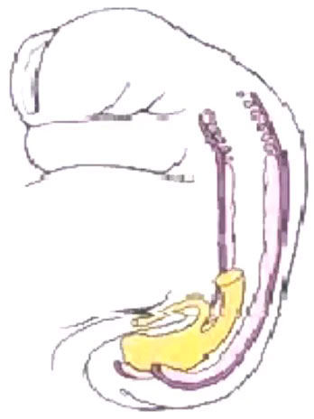
Male Human Embryo (week 8, stage 22) Detail from cross-section showing mesonephros, mesonephric duct, paramesonephric duct and developing testis. |
Until the end of the 6th week the male and female genital systems are indistinguishable. Sex differentiation is based upon the presence of the specific sex chromosomes.
|
Male

|

|

|
| peritoneal cavity | rete tesits | G3 testis |

- week 8 - males Sertoli cells secrete anti müllerian hormone (AMH), which causes regression of the paramesonephric ducts between the 8th and 10th weeks.
- week 9 to 10 - gonadal cells begin to produce testosterone, which maintains the mesonephric ducts.
- mesonephric ducts go on to form the internal genital tract:
- rete testis
- ductuli efferentes
- vas deferens
Female

- during the same time course as above, the opposite occurs.
- The sex cords degenerate and the genital ridge forms secondary cortical sex cords.
- These induce the primordial germ cells to form the ovarian follicles.
- Due to the lack of AMH and testosterone, the mesonephric ducts degenerate
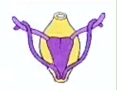
Paramesonephric (Müllerian) ducts form the internal female genital tract
In both sexes, the external genitalia appear similar until week 12 (GA week 14).
Trigone
| <mediaplayer width='445' height='365' image="http://php.med.unsw.edu.au/embryology/images/5/5b/Trigone_001_icon.jpg">File:Trigone_001.mp4</mediaplayer> | This animation shows the posterior of the developing bladder between Week 4 and 6.
|
Additional Information
| Additional Information - Content shown under this heading is not part of the material covered in this class. It is provided for those students who would like to know about some concepts or current research in topics related to the current class page. |
References
BGDB: Lecture - Gastrointestinal System | Practical - Gastrointestinal System | Lecture - Face and Ear | Practical - Face and Ear | Lecture - Endocrine | Lecture - Sexual Differentiation | Practical - Sexual Differentiation | Tutorial
Glossary Links
- Glossary: A | B | C | D | E | F | G | H | I | J | K | L | M | N | O | P | Q | R | S | T | U | V | W | X | Y | Z | Numbers | Symbols | Term Link
Cite this page: Hill, M.A. (2024, May 21) Embryology BGDB Sexual Differentiation - Late Embryo. Retrieved from https://embryology.med.unsw.edu.au/embryology/index.php/BGDB_Sexual_Differentiation_-_Late_Embryo
- © Dr Mark Hill 2024, UNSW Embryology ISBN: 978 0 7334 2609 4 - UNSW CRICOS Provider Code No. 00098G