BGDB Practical - Upper Gastrointestinal Tract Histology
Introduction

This current page provides background support information for Medicine phase 1 BGD Histology Practical Virtual Slides. Page does not form part of the BGDB practical class virtual slides.
- Virtual Slides: Upper Gastrointestinal Tract Histology (requires zpass login)
Practical 4: Upper Gastrointestinal Tract Histology Principal Teacher: Patrick de Permentier
Aim
To introduce the histology of the upper GIT (gastro-intestinal tract) Specific Objectives:
- To describe the histology of the lip and the tongue, including epithelia, muscles, glands, papillae and taste buds.
- To appreciate the histological features of the three major salivary glands.
- To describe the general architecture of the wall of the alimentary canal and the functions of the mucosa, submucosa, muscle layer and serosal or adventitial layer in the various anatomical divisions of the canal.
- To describe the main features of the structure of the wall of the oesophagus, cardio-oesophageal junction, stomach, and duodenum.
- To identify the cells of gastric epithelium and distinguish the histological features of the cardiac, body and pyloric regions of stomach. To appreciate the role of mucus on the surface of gastric epithelium.
- To describe the histological features of the duodenum including villi, intestinal mucosal glands (crypts of Lieberkühn), lymphatic tissue and nerve plexuses.
Learning Activities
Lip and Infant lip
In these 2 slides, identify the mucosal surface and skin surface on opposite sides of the lip. Note the mucocutaneous transition zone (also called "red margin" or vermillion border). Observe the orbicularis oris skeletal muscle; labial salivary glands (mucoserous glands; mostly serous in infant lip); salivary ducts with stratified epithelium; nerve fascicles.
Soft Palate
What type of epithelium covers the respiratory and oral surface respectively? Note palatal salivary glands secreting mucus; ducts lined by stratified cuboidal epithelium; skeletal muscle; palatine tonsil.
Root of Tongue
It shows interlacing striated muscle fibres, lingual tonsils with lymphatic nodules, crypts, mucoserous glands, and ducts of the glands (some mucous acini have serous demilunes). Tongue: circumvallate papillae Taste buds in the lateral wall of circumvallate papillae; von Ebner's serous glands and ducts; striated muscle fascicles; blood vessels; nerve fascicles; lymph aggregations.
Tongue (rabbit)
Foliate papillae with taste buds in walls; serous glands; LS and TS of striated muscle fibres.

Submandibular gland
Mixed salivary gland; predominantly serous acini; some mucous acini with serous demilunes; interlobular ducts with stratified cuboidal or stratified columnar epithelium; connective tissue; striated ducts with simple cuboidal lining epithelium; short intercalated ducts.
Sublingual gland
Predominantly mucous acini; some serous demilunes; interlobular ducts with stratified cuboidal/columnar epithelium; connective tissue; striated ducts with simple columnar lining epithelium; short intercalated ducts.


Parotid gland
Serous salivary gland; lobules; connective tissue septa; serous acini, zymogen granules; striated ducts; interlobular ducts with stratified epithelium; intercalated ducts; lymph node with capsule.


Oesophagus
Mucosa; submucosa; muscularis externa.
- What type of epithelium lines the lumen?
- What types of muscle fibres can you recognize in the 2 sublayers of the muscularis externa?


Cardio-oesophageal junction

Oesophageal epithelium; sharp transition to gastric epithelium; cardiac glands (of stomach); smooth muscle fascicles.
- What are the 2 sublayers of the muscularis externa in the oesophagus?
stomach labeled overview
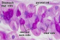
parietal cells - chief cells
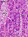
mucus neck - parietal cells - chief cells
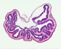
stomach overview
stomach mucosa
mucosa - secretory epithelial sheath - goblet cell
gastric glands - parietal cells - chief cells
stomach overview


- Stomach Histology Links: stomach labeled overview | parietal cells - chief cells | mucus neck - parietal cells - chief cells | stomach overview | stomach mucosa | mucosa - secretory epithelial sheath - goblet cell | gastric glands - parietal cells - chief cells | stomach overview | Stomach Histology | Stomach Development | Gastrointestinal Tract Development
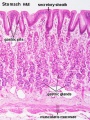
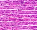
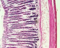
Stomach fundus
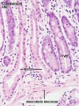
Surface secretory sheet gland (secreting mucus) folded macroscopically to form gastric rugae; gastric pits; gastric simple branched tubular glands; mucous neck cells; parietal cells (pale staining); zymogen (chief) cells (dark staining) in the deeper regions of glands; muscularis mucosae; extensive submucosa; muscularis externa (part).
Pyloro-duodenal junction
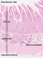
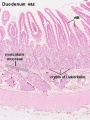
In the pyloric region of stomach, note the deep pyloric pits and shorter, straighter pyloric glands lined mainly by mucus-secreting cells (the specimen shows some post-mortem loss of mucins). Note the pyloric sphincter (smooth muscle) and the transition of epithelium at the duodenum with its villi and intestinal crypts (of Lieberkühn). A striking feature of the first part of the duodenum is the presence of Brunner's submucosal glands.
- What is the secretion from Brunner’s glands?
- What structure defines the location of Brunner’s glands as being part of the submucosa?

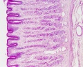
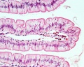
In the duodenum of the small intestine, surface epithelium of columnar absorptive cells; goblet cells; villi; intestinal crypts (tubular glands); duodenal (Brunner's) glands of submucosa; submucosa; muscularis externa with two sublayers; lacteals. This part of the duodenum is retroperitoneal and so it is covered by a tunica adventitia and not a tunica serosa. (Paneth cells in the bases of the crypts of Lieberkühn are not stained in this preparation.)
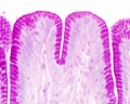
Duodenum overview
Duodenum villi and crypts
Duodenum
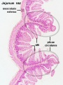
Jejunum overview
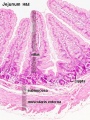
Jejunum villus
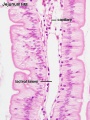
Jejunum labeled
Jejunum unlabeled


