BGDB Face and Ear - Late Embryo: Difference between revisions
mNo edit summary |
mNo edit summary |
||
| Line 94: | Line 94: | ||
{| | {| | ||
| [[File:Gray0924.jpg| | | [[File:Gray0924.jpg|500px]] | ||
Membranous Labrynth | Adult Membranous Labrynth | ||
| | | | ||
===The Membranous Labyrinth=== | ===The Membranous Labyrinth=== | ||
The membranous labyrinth is lodged within the bony cavities just described, and has the same general form as these; it is, however, considerably smaller, and is partly separated from the bony walls by a quantity of fluid, the perilymph. In certain places it is fixed to the walls of the cavity. The membranous labyrinth contains fluid, the endolymph, and on its walls the ramifications of the acoustic nerve are distributed. | |||
Within the osseous vestibule the membranous labyrinth does not quite preserve the form of the bony cavity, but consists of two membranous sacs, the utricle, and the saccule. | Within the osseous vestibule the membranous labyrinth does not quite preserve the form of the bony cavity, but consists of two membranous sacs, the utricle, and the saccule. | ||
===The Utricle=== | ===The Utricle=== | ||
The utricle, the larger of the two, is of an oblong form, compressed transversely, and occupies the upper and back part of the vestibule, lying in contact with the recessus ellipticus and the part below it. That portion which is lodged in the recess forms a sort of pouch or cul-de-sac, the floor and anterior wall of which are thickened, and form the macula acustica utriculi, which receives the utricular filaments of the acoustic nerve. The cavity of the utricle communicates behind with the semicircular ducts by five orifices. From its anterior wall is given off the ductus utriculosaccularis, which opens into the ductus endolymphaticus. | |||
===The Saccule=== | ===The Saccule=== | ||
The saccule is the smaller of the two vestibular sacs; it is globular in form, and lies in the recessus sphæricus near the opening of the scala vestibuli of the cochlea. Its anterior part exhibits an oval thickening, the macula acustica sacculi, to which are distributed the saccular filaments of the acoustic nerve. Its cavity does not directly communicate with that of the utricle. From the posterior wall a canal, the ductus endolymphaticus, is given off; this duct is joined by the ductus utriculosaccularis, and then passes along the aquæductus vestibuli and ends in a blind pouch (saccus endolymphaticus) on the posterior surface of the petrous portion of the temporal bone, where it is in contact with the dura mater. From the lower part of the saccule a short tube, the canalis reuniens of Hensen, passes downward and opens into the ductus cochlearis near its vestibular extremity. | |||
===The Semicircular Ducts=== | ===The Semicircular Ducts=== | ||
(''ductus semicirculares''; membranous semicircular canals), The semicircular ducts are about one-fourth of the diameter of the osseous canals, but in number, shape, and general form they are precisely similar, and each presents at one end an ampulla. They open by five orifices into the utricle, one opening being common to the medial end of the superior and the upper end of the posterior duct. In the ampullæ the wall is thickened, and projects into the cavity as a fiddle-shaped, transversely placed elevation, the septum transversum, in which the nerves end. | (''ductus semicirculares''; membranous semicircular canals), The semicircular ducts are about one-fourth of the diameter of the osseous canals, but in number, shape, and general form they are precisely similar, and each presents at one end an ampulla. They open by five orifices into the utricle, one opening being common to the medial end of the superior and the upper end of the posterior duct. In the ampullæ the wall is thickened, and projects into the cavity as a fiddle-shaped, transversely placed elevation, the septum transversum, in which the nerves end. | ||
| Line 114: | Line 111: | ||
The utricle, saccule, and semicircular ducts are held in position by numerous fibrous bands which stretch across the space between them and the bony walls. | The utricle, saccule, and semicircular ducts are held in position by numerous fibrous bands which stretch across the space between them and the bony walls. | ||
|} | |} | ||
{{BGDBFooter}} | {{BGDBFooter}} | ||
Revision as of 09:51, 16 May 2017
Week 6
Primary Palate
- Beginning week 6 there is fusion of the upper lip.
- Formed by the maxillary prominences of of the first pharyngeal arch and the frontonasal prominence.
- Failure of this embryonic process leads to cleft lip.
Week 6 (stage 16)
Week 6 (stage 17)
Week 7 (stage 18)
Week 7 (stage 19)


Above images show face development through week 6 to week 7 (1mm scale markings).
The animation shows the early fusion of the primary palate in the human embryo between stage 17 and 18, going from an epithelial seam to the mesenchymal bridge.
 Face Development Movie | MP4 movie
|
This animation shows a ventral view of development of the human face from approximately week 5 through to neonate.
|
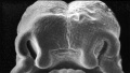
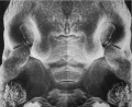
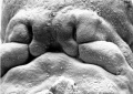
Week 8
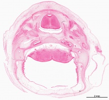
- Oral Cavity (stage 23)
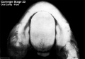
Floor
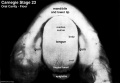
Floor (labeled)

Roof
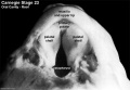
Roof (labeled)


Selected Head Images: B4 - Choroid Plexus | B5 - Cochlea | B6 - Cochlea
Palate

|
The dark "pear-shaped" central structure at the top is the developing tongue. The two pale regions either side are the palatal shelves, note that they have not yet fused in the midline (failure of this process is cleft palate). |
Hearing
|
|
Behind that a pale cartilagenous region (that later ossifies) encloses the structuctures of the inner ear, beside which middle ear bones are forming. On the righthand side of the head the external ear is visible. The lower half of the image shows the developing brainstem with a large ventricular space occupied in part by an extensive choroid plexus (manufacturer of cerebrospinal fluid). |
|

Embryonic development of the human membranous labyrinth. |

Membranous labyrinth and acoustic complex model reconstructed from week 8 human embryo (CRL 30 mm). Note that by the end of the embryonic period the adult external structure has been achieved, internal development continues through the fetal period. |
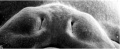
Embryonic External Ear
Shown below are the changes in external ear development between week 5 to week 8. Development changes from a series of 6 hillocks on arch 1 and arch 2 (week 5) to a structure resembling the adult ear (week 8).

Additional Information
| Additional Information - Content shown under this heading is not part of the material covered in this class. It is provided for those students who would like to know about some concepts or current research in topics related to the current class page. |
Historic
Streeter GL. On the development of the membranous labyrinth and the acoustic and facial nerves in the human embryo. (1906) Amer. J Anat. 6:139-165.

Adult Membranous Labrynth |
The Membranous LabyrinthThe membranous labyrinth is lodged within the bony cavities just described, and has the same general form as these; it is, however, considerably smaller, and is partly separated from the bony walls by a quantity of fluid, the perilymph. In certain places it is fixed to the walls of the cavity. The membranous labyrinth contains fluid, the endolymph, and on its walls the ramifications of the acoustic nerve are distributed. Within the osseous vestibule the membranous labyrinth does not quite preserve the form of the bony cavity, but consists of two membranous sacs, the utricle, and the saccule. The UtricleThe utricle, the larger of the two, is of an oblong form, compressed transversely, and occupies the upper and back part of the vestibule, lying in contact with the recessus ellipticus and the part below it. That portion which is lodged in the recess forms a sort of pouch or cul-de-sac, the floor and anterior wall of which are thickened, and form the macula acustica utriculi, which receives the utricular filaments of the acoustic nerve. The cavity of the utricle communicates behind with the semicircular ducts by five orifices. From its anterior wall is given off the ductus utriculosaccularis, which opens into the ductus endolymphaticus. The SacculeThe saccule is the smaller of the two vestibular sacs; it is globular in form, and lies in the recessus sphæricus near the opening of the scala vestibuli of the cochlea. Its anterior part exhibits an oval thickening, the macula acustica sacculi, to which are distributed the saccular filaments of the acoustic nerve. Its cavity does not directly communicate with that of the utricle. From the posterior wall a canal, the ductus endolymphaticus, is given off; this duct is joined by the ductus utriculosaccularis, and then passes along the aquæductus vestibuli and ends in a blind pouch (saccus endolymphaticus) on the posterior surface of the petrous portion of the temporal bone, where it is in contact with the dura mater. From the lower part of the saccule a short tube, the canalis reuniens of Hensen, passes downward and opens into the ductus cochlearis near its vestibular extremity. The Semicircular Ducts(ductus semicirculares; membranous semicircular canals), The semicircular ducts are about one-fourth of the diameter of the osseous canals, but in number, shape, and general form they are precisely similar, and each presents at one end an ampulla. They open by five orifices into the utricle, one opening being common to the medial end of the superior and the upper end of the posterior duct. In the ampullæ the wall is thickened, and projects into the cavity as a fiddle-shaped, transversely placed elevation, the septum transversum, in which the nerves end. The utricle, saccule, and semicircular ducts are held in position by numerous fibrous bands which stretch across the space between them and the bony walls. |
BGDB: Lecture - Gastrointestinal System | Practical - Gastrointestinal System | Lecture - Face and Ear | Practical - Face and Ear | Lecture - Endocrine | Lecture - Sexual Differentiation | Practical - Sexual Differentiation | Tutorial
Glossary Links
- Glossary: A | B | C | D | E | F | G | H | I | J | K | L | M | N | O | P | Q | R | S | T | U | V | W | X | Y | Z | Numbers | Symbols | Term Link
Cite this page: Hill, M.A. (2024, April 26) Embryology BGDB Face and Ear - Late Embryo. Retrieved from https://embryology.med.unsw.edu.au/embryology/index.php/BGDB_Face_and_Ear_-_Late_Embryo
- © Dr Mark Hill 2024, UNSW Embryology ISBN: 978 0 7334 2609 4 - UNSW CRICOS Provider Code No. 00098G