|
|
| (42 intermediate revisions by 2 users not shown) |
| Line 1: |
Line 1: |
| {{Header}}
| | ==Organogenesis Lab== |
| =Muscle Development = | |
|
| |
|
| == Introduction ==
| | Please note the different location for this week’s practical class: Wallace Wurth Teaching Lab 116. |
| [[File:Skeletal muscle structure cartoon.jpg|thumb|Skeletal Muscle]]
| |
| This laboratory concerns the development and differentiation of skeletal muscle, fibre type differentiation, muscle stem cells and muscle disease.
| |
|
| |
|
| The histology of skeletal muscle has been covered in ANAT2241.
| |
|
| |
|
| | '''PRACTICAL CLASS PROGRAM''' |
|
| |
|
| ==Objectives==
| | * Weekly Quiz + revision (15 minutes) |
| | * Practical class activities (90 minutes) |
| | * Practical Class Revision (15 minutes) |
|
| |
|
| # Understand the origin, differentiation and development of skeletal muscle tissue.
| |
| # Know what is meant by patterning, conversion and adult plasticity of muscle fibre type.
| |
| # Develop an understanding of research methods for studying skeletal muscle abnormalities.
| |
|
| |
|
| ==Muscle Contraction==
| | '''PRACTICAL CLASS ACTIVITIES (90 minutes)''' |
| {|
| |
| ! Crossbridge animation
| |
| ! Component proteins
| |
| |-
| |
| | [[File:Actin_myosin_crossbridge_3D_animation.gif|link=File:Actin myosin crossbridge 3D animation.gif]]
| |
| | [[File:Actin_myosin_crossbridge_3D_animation.jpg|link=File:Actin myosin crossbridge 3D animation.gif]]
| |
| |-
| |
| ! Sarcomere Animation
| |
| ! Muscle Histology
| |
| |-
| |
| | [[File:Sarcomere_animation.gif]]
| |
| | [[File:Skeletal muscle histology 016.jpg|350px]]
| |
| |}
| |
|
| |
|
| ==Sarcomere - Electron Microscopy==
| | * Fertile egg dissections, stage definition, and annotations of structures (first 60 minutes) |
| {|
| | * Observation of skeletal preparations of chicken and mouse foetuses (first 60 minutes) |
| ! Virtual Slides
| | * Group presentation of annotated embryo images (final 30 minutes) |
| |-
| |
| | valign=bottom|{{SlideSkeletalMuscleEM01}}
| |
| | valign=bottom|{{SlideSkeletalMuscleEM02}}
| |
| | valign=bottom|{{SlideSkeletalMuscleEM03}}
| |
| |-
| |
| |}
| |
|
| |
|
| [http://www.lab.anhb.uwa.edu.au/mb140/CorePages/Muscle/Muscle.htm#SKELETAL Skeletal Muscle Histology]
| |
|
| |
|
| | '''LEARNING OBJECTIVES''' |
|
| |
|
| ==Muscle Fibre Types==
| | * Understanding early neurulation, mesoderm and heart development, and being able to identify the defining structures in the chicken embryo. |
| {|
| | * Understanding craniofacial and limb development and being able to identify the defining structures in chicken embryos. |
| | width=410px|[[File:Muscle fiber types.jpg]]
| | * Understanding the development of the musculoskeletal system and being able to identify the defining structures in chicken embryos. |
| | '''Type I fibres'''
| | * Be able to apply basic practical laboratory skills and work with embryo and regeneration models. |
| * Red muscles contain predominantly (but not exclusively) red muscle cells. Red muscle fibres are comparatively thin and contain large amounts of myoglobin and mitochondria. | | * Be able to work effectively within a small team to complete academic tasks. |
| * Red fibres contain an isoform of myosin with low ATPase activity, i.e. the speed with which myosin is able to use up ATP. Contraction is therefore '''slow'''. | | * Be able to present embryonic observations effectively and appropriately to an audience |
| * Red muscles are used when sustained production of force is necessary, e.g. in the control of posture. | | * Be able to self-manage and work independently with an ability to take responsibility for their own learning, and an appreciation of the value of learning. |
|
| |
|
| '''Type II fibres'''
| |
| * White muscle cells, which are predominantly found in white muscles, are thicker and contain less myoglobin. ATPase activity of the myosin isoform in white fibres is high, and contraction is '''fast'''.
| |
| ** '''Type IIA''' fibres (red) contain many mitochondria and are available for both sustained activity and short-lasting, intense contractions.
| |
| ** '''Type IIB/IIX''' fibres (white) contain only few mitochondria. They are recruited in the case of rapid accelerations and short lasting maximal contraction. Type IIB/IIX fibres rely on anaerobic glycolysis to generate the ATP needed for contraction.
| |
| |}
| |
|
| |
|
|
| |
|
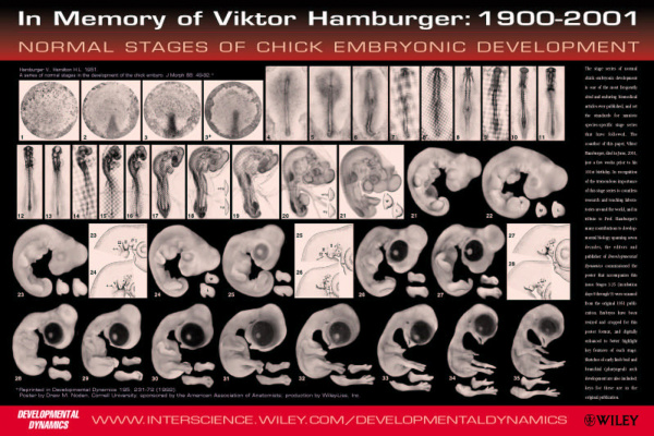
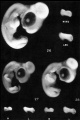
| ==Muscle Embryology== | | [[File:Chicken_Embryo_Hamburger_stages.jpg|600px|link=Hamburger Hamilton Stages]] |
| * Determined - when cells are located within the somite myotome.
| |
| * Migrate - small cell population moves to location of adult muscle.
| |
| * Proliferate - as myoblasts, single nuclei cells.
| |
| * Fuse - to form myotubes, the primitive skeletal muscle fibre not yet showing sarcomeres.
| |
| * Differentiate - express contractile proteins
| |
|
| |
|
| | ''These are the Hamburger stages of chicken development'' |
|
| |
|
| ==Muscle Stem Cells==
| |
|
| |
|
| {|
| | See also the [https://www.jove.com/video/306/windowing-chicken-eggs-for-developmental-studies JoVE article on chicken egg preparation]: <pubmed>18989413</pubmed> |
| ! Satellite cells
| |
| |-
| |
| | These cells remain as muscle stem cells and lie under the basal lamina around each skeletal muscle fibre.
| |
|
| |
|
| They have a role in postnatal growth and also regeneration of muscle fibres.
| |
|
| |
|
| This stem cell population is able to differentiate and replace damaged muscle fibres.
| | ===Additional Chicken Links=== |
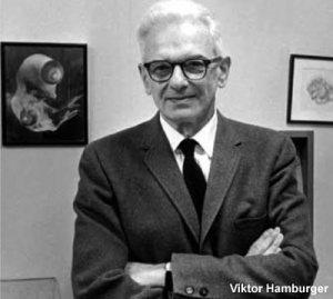
| | [[File:Viktor Hamburger.jpg|thumb|alt=Viktor Hamburger|link=Embryology History - Viktor Hamburger|Viktor Hamburger (1900 – 2001)]] |
| | More about chicken embryogenesis: [[Chicken Development]] | [[Hamburger Hamilton Stages]] |
| | <br> |
| | {{Chicken links}} |
| | <br> |
|
| |
|
| {| class="wikitable mw-collapsible mw-collapsed"
| | <gallery> |
| ! More recent papers
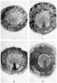
| | File:HHstage1-4.jpg|stage 1-4 |
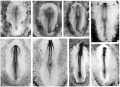
| |- | | File:HHstage5-10.jpg|stages 5-10 |
| | [[File:Mark_Hill.jpg|90px|left]] {{Most_Recent_Refs}} | | File:HHstage11-14.jpg|stages 11-14 |
| | File:HHstage15-18.jpg|stages 15-18 |
| | File:HHstage19-21.jpg|stages 19-21 |
| | File:HHstage22-25.jpg|stages 22-25 |
| | File:HHstage26-28.jpg|stages 26-28 |
| | File:HHstage29-32.jpg|stage 29-32 |
| | </gallery> |
|
| |
|
| Search term: [http://www.ncbi.nlm.nih.gov/pubmed/?term=Satellite+Cell ''Satellite Cell'']
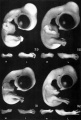
| | [[File:Mouse_vs_Human_embryogenesis.jpg]] |
|
| |
|
| <pubmed limit=5>Satellite Cell</pubmed>
| | ''This figure compares the human and mouse developmental stages'' |
| |}
| |
|
| |
|
| :'''Links:''' [[Stem Cells]] | | More about Mouse embryogenesis: [[Mouse Timeline Detailed]] |
| | [[File:Muscle satellite cell EM02.jpg|300px]]
| |
| |}
| |
|
| |
|
| ==Muscular Dystrophies==
| |
| [[File:X-Linked recessive (carrier mother).jpg|thumb|X-linked recessive (carrier mother)]]
| |
| Dystrophy refers to the degeneration of a tissue, muscular dystrophy is degeneration of skeletal muscle.
| |
|
| |
|
| * '''Duchenne Muscular Dystrophy''' - (DMD) The most common occuring in Boys and in Duchenne Muscular Dystrophy (DMD).
| | {{Chicken}} |
| * '''Autosomal Recessive Muscular Dystrophy''' - Dystroglycan, a protein that associates with both dystrophin and membrane molecules, is a candidate gene for the site of the mutation in autosomal recessive muscular dystrophies. A knockout mouse has been generated that has early developmental abnormalities.
| |
| * '''Myotonic Dystrophy''' - An inherited disorder in which the muscles contract but have decreasing power to relax. With this condition, the muscles also become weak and waste away. The myotonic dystrophy gene, found on chromosome 19, codes for a protein kinase that is found in skeletal muscle, where it likely plays a regulatory role. The disease is "amplified" through generations probably by a similar GC expansion associated with Huntington disease.
| |
| * '''Facioscapulohumeral Muscular Dystrophy''' - (FSHD) This form of muscular dystrophy is an autosomal dominant genetic disorder characterized by progressive muscle weakness and wasting that typically begins in the face, shoulder-girdle and upper arm. Currently thought to relate to an impaired muscle regeneration process.
| |
|
| |
|
| | | ===External Links=== |
| | |
| == Lab 7 Assessment Questions== | |
| | |
| | |
| TBA
| |
| | |
| | |
| ===Search ===
| |
| | |
| * '''Bookshelf''' [http://www.ncbi.nlm.nih.gov/sites/entrez?db=Books&cmd=search&term=muscle_development muscle development] | [http://www.ncbi.nlm.nih.gov/sites/entrez?db=Books&cmd=search&term=muscle_fiber_type muscle fiber type] | [http://www.ncbi.nlm.nih.gov/sites/entrez?db=Books&cmd=search&term=troponin troponin]
| |
| | |
| * '''Pubmed''' [http://www.ncbi.nlm.nih.gov/sites/gquery?itool=toolbar&cmd=search&term=muscle_fiber_type muscle fiber type] | [http://www.ncbi.nlm.nih.gov/sites/gquery?itool=toolbar&cmd=search&term=troponin troponin] | [http://www.ncbi.nlm.nih.gov/sites/gquery?itool=toolbar&cmd=search&term=Gtf2ird1 Gtf2ird1] |
| |
| | |
| | |
| | |
| | |
| == Terms ==
| |
| | |
| '''anterior tibialis''' - (tibialis anterior) skeletal muscle situated on the lateral side of the tibia and is a direct flexor of the foot at the ankle-joint.
| |
| | |
| '''carrier females''' - (carrier mother, carrier) X-linked recessive inheritance seen with DMD and a range of other inherited disorders where the gene is recessive and located on the X chromosome.
| |
| | |
| '''cis-acting elements''' - DNA sequences that through transcription factors or other trans-acting elements or factors, regulate the expression of genes on the same chromosome.
| |
| | |
| '''corticosteroids''' - used as a pharmacologic agents in DMD treatment these are the synthetic glucocorticoids; prednisone and its active metabolite prednisolone and
| |
| deflazacort an inactive prodrug.
| |
| | |
| '''enhancer''' - A cis-regulatory sequence that can regulate levels of transcription from an adjacent promoter. Many tissue-specific enhancers can determine spatial patterns of gene expression in higher eukaryotes. Enhancers can act on promoters over many tens of kilobases of DNA and can be 5' or 3' to the promoter they regulate.
| |
| | |
| '''extensor digitorum longus''' - (EDL) skeletal muscle situated at the lateral part of the front of the leg and extend the phalanges of the toes, and, continuing their action, flex the foot upon the leg.
| |
| | |
| '''Gtf2ird1''' - General Transcription Factor 2 -I Repeat domain-containing protein 1. [http://www.ncbi.nlm.nih.gov/entrez/dispomim.cgi?id=604318 OMIM: Gtf2ird1]
| |
| | |
| '''MusTRD1''' - muscle TFII-I repeat domain-containing protein 1.
| |
| | |
| '''MyHC''' - acronym for myosin heavy chain.
| |
| | |
| '''myoblast''' - the undifferentiated mononucleated muscle cell progenitor, which in skeletal muscle fuses to form a myotube, that in turn expresses contractile proteins to form a muscle fibre.
| |
| | |
| '''myosin heavy chain''' - protein forming the thick filament of the sarcomere and the motor for actin-myosin contraction. There are 17 different myosin classes.
| |
| | |
| '''myotube''' - the initial multinucleated cell formed by fusion of myoblasts during skeletal muscle development.
| |
| | |
| '''promoter''' - A regulatory region a short distance upstream from the 5' end of a transcription start site that acts as the binding site for RNA polymerase II. A region of DNA to which RNA polymerase IIbinds in order to initiate transcription.
| |
| | |
| '''regulatory sequence''' - (regulatory region, regulatory area) is a segment of DNA where regulatory proteins such as transcription factors bind preferentially.
| |
| | |
| '''soleus''' - slow skeletal muscle situated immediately in front of the gastrocnemius and when standing taking its fixed point from below, steadies the leg upon the foot and prevents the body from falling forward.
| |
| | |
| '''Troponin''' - striated muscle contraction is regulated by the calcium-ion-sensitive, multiprotein complex troponin and the fibrous protein tropomyosin. Troponin has 3 subunits (TnC, TnI, TnT) and is located on the actin filament. [http://www.ncbi.nlm.nih.gov/entrez/dispomim.cgi?id=191040 OMIM: Troponin I slow]
| |
| | |
| | |
| == External Links == | |
| {{External Links}} | | {{External Links}} |
|
| |
|
| * Blue Histology [http://www.lab.anhb.uwa.edu.au/mb140/CorePages/Muscle/Muscle.htm#SKELETAL Skeletal Muscle histology] | | * JOVE - [http://www.jove.com/science-education/5153/an-introduction-to-the-chick-gallus-gallus-domesticus An Introduction to the Chicken] |
| * [http://currents.plos.org/md/ PLOS Currents - Muscular Dystrophy]
| |
| * [http://omim.org/entry/310200 OMIM]
| |
| * Primer [http://www.plosbiology.org/article/info%3Adoi%2F10.1371%2Fjournal.pbio.0020348 Skeletal Muscle Fiber Type: Influence on Contractile and Metabolic Properties]
| |
| | |
| | |
| {{Glossary}}
| |
| | |
| {{Footer}}
| |
| | |
| | |
| ==References==
| |
| | |
| <references/>
| |
| | |
| {{2016ANAT2341}}
| |


