2010 Group Project 1: Difference between revisions
No edit summary |
|||
| Line 72: | Line 72: | ||
===3D Ultrasound=== | ===3D Ultrasound=== | ||
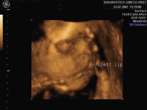
[[File:Z3DCleft_Lip_Picture.jpg|thumb|right|3D ultrasound image showing a fetus with a cleft lip abnormality]] | |||
Three-dimensional ultrasound images are produced in one of several ways. The first is to have several arrays of 2D transducers working together to collect a series of 2D cross-sections and combine these into a 3D image, though this can be difficult. 3D ultrasound images can also be made with the use of arrays of 1D transducers to create 2D B-scan images of known areas in 3D space. The 1D arrays can either by swept across the patient by machine or by the technician. 3D ultrasound images are particularly useful for visualising the volumes of structures, and better viewing complex 3D structures. However, even today it is mostly 2D ultrasound that is used, with 3D ultrasound only used in select applications. <ref>Kremkali, F.W. (2006) Diagnostic Ultrasound Principles and Instruments (7th ed.) St Louis: Saunders Elsevier. pp. 6</ref><ref><pubmed>20349815</pubmed></ref> | Three-dimensional ultrasound images are produced in one of several ways. The first is to have several arrays of 2D transducers working together to collect a series of 2D cross-sections and combine these into a 3D image, though this can be difficult. 3D ultrasound images can also be made with the use of arrays of 1D transducers to create 2D B-scan images of known areas in 3D space. The 1D arrays can either by swept across the patient by machine or by the technician. 3D ultrasound images are particularly useful for visualising the volumes of structures, and better viewing complex 3D structures. However, even today it is mostly 2D ultrasound that is used, with 3D ultrasound only used in select applications. <ref>Kremkali, F.W. (2006) Diagnostic Ultrasound Principles and Instruments (7th ed.) St Louis: Saunders Elsevier. pp. 6</ref><ref><pubmed>20349815</pubmed></ref> | ||
Revision as of 18:46, 10 September 2010
Ultrasound
Introduction
History
How It Works
The basic principle of ultrasound is similar to that of sonar – that is, sending out pulses of high-frequency sound waves, receiving back the echoes of those waves after they bounce off surrounding materials, and constructing a picture of those materials from the received sounds. In medical ultrasound, or sonography, pulses of ultrasound are repeatedly sent into the body where they bounce off the edges of organs and tissues, relaying that anatomical information back to the ultrasound machine. [1]
There are three main elements in an ultrasound system: the transducer, the machine and the recording devices [2].
The Transducer
The transducer is the piece of equipment in contact with the patient. It emits the ultrasound pulses and receives the echoes of those pulses. Information about the patient’s tissues is generated by calculating the time it takes for the ultrasound pulses to scatter and bounce off those tissues and travel back to the transducer. The ultrasound waves will take longer or shorter amounts of time to travel through different tissues depending on their properties.
Since air diminishes the integrity of the ultrasound signals (the ultrasound can bounce off even a thin layer of air, almost completely preventing the penetration of the ultrasound into the patient’s tissues), a medium such as a water-soluble gel is applied between the transducer and the patient’s skin to improve the interface between the two. There are several kinds of transducers producing different kinds of ultrasound images, discussed below. Transducers can be non-invasive (for prenatal diagnosis, pressed against the woman's pelvis or abdomen) or they can be invasive (transvaginal transducers, for example, are inserted into the vagina to obtain better views of the ovaries, fallopian tubes, uterus, and surrounding structures).
Within the transducer are elements made of silicon crystals. The pulses of ultrasound are created using the piezoelectric effect – when silicon crystals experience electrical impulses they mechanically deform on a microscopic level, producing high-frequency sound waves. Returning sound waves (echoes) will similarly deform the silicon crystals, and these deformations are converted to electrical impulses which are then sent to the ultrasound machine for processing. Different kinds of transducers will produce different kinds of images, or scans. High-frequency transducers (5MHz or greater) give better resolution of images, but low frequency transducers penetrate deeper into tissues and so provide images of deeper structures. [3]
Sending one ultrasound pulse into the body will generate a line of dots – this represents one line of echo information, or one scan line. (DIAGRAM) Not all of that pulse bounces back off one single anatomical structure; instead, most of the pulse continues through to deeper structures where some of the pulse bounces back from each structure it passes through. This is what generates the line of dots representing an internal view of the patient. Since the echoes of a single pulse generate one scan line of information, generating several scan lines next to one another in an ordered sequence will generate a cross-sectional image of the patient. This way of directing pulses through the tissues is termed “scanning” or “sweeping”. Scanning is performed quickly and automatically by an “array” of organised group of transducer elements, creating many still images or frames which are viewed in sequence, like a movie. Thus, ultrasound exams are performed in real-time. [4]
Different transducers have different arrays of piezoelectric elements, and produce a variety of different scans or images:
| Linear Array Transducers | Curved or Convex Array Transducers | Phased Array Transducers |
|---|---|---|
| Elements are arranged in a straight line, firing pulses vertically and parallel to each other | Elements are arranged nest to each other in a convex shape, emitting pulses in a sunburst-like manner | Emit pulses from a compact line of elements in slightly different directions in rapid sequence |
| Produces a wide-view linear or rectangular image | Produces a fan-shaped sector scan with a concave top border | Produces a sector scan shaped like a slice of pizza |
| Image goes here | Image goes here | Image goes here |
The Ultrasound Machine and Recording Devices
The ultrasound machine itself is comprised of the computer hardware and software needed to convert the signals picked up by the transducer into an image that can be viewed. The image is in greyscale, using various shades from black to white to indicate different tissues and tissue features (ILLUSTRATIVE PICTURE.)
The other basic part of the ultrasound system is the recording equipment – that is, a range of devices such as multiformat cameras, video printers and video recorders used to store information from the patient’s examination.
So far, what has been explained are the basic principles of how a normal 2D ultrasound image is generated. There have been are, however, some developments in ultrasound technology that allow the generation of slightly different images to the normal 2D ones, providing more diagnostic information. These are the Doppler ultrasound and 3D ultrasound imaging.
Doppler Ultrasound
Doppler ultrasound utilises the Doppler effect to view moving structures, such as the flow of blood through major blood vessels of the heart. The Doppler effect refers to the change in frequency of a wave relative to the motion of the wave source and the observer. In a Doppler ultrasound, sound waved are reflected from moving structures with a shift in frequency of those sound waves proportional to the velocity of the moving structure.[5]
Continuous-wave Doppler ultrasound detects signals from anything moving in the path of the pulse, and cannot distinguish well between separate vessels. Pulsed Doppler ultrasound uses a pulse in a thin, focused beam and detects signals from only one point in that beam, allowing the imaging of specific vessels. Colour Doppler imaging is a type of pulsed Doppler ultrasound where software is used to overlay the greyscale image with colours representing frequency shifts. Different colours are assigned to indicate movement towards or away from the transducer. [6][7]
Link to more ultrasound images | Ultrasound Images
Link to video of the Doppler Effect | Assignment Discovery: the Doppler Effect
3D Ultrasound
Three-dimensional ultrasound images are produced in one of several ways. The first is to have several arrays of 2D transducers working together to collect a series of 2D cross-sections and combine these into a 3D image, though this can be difficult. 3D ultrasound images can also be made with the use of arrays of 1D transducers to create 2D B-scan images of known areas in 3D space. The 1D arrays can either by swept across the patient by machine or by the technician. 3D ultrasound images are particularly useful for visualising the volumes of structures, and better viewing complex 3D structures. However, even today it is mostly 2D ultrasound that is used, with 3D ultrasound only used in select applications. [8][9]
Link to more ultrasound images | Ultrasound Images
Link to a review of 3D ultrasound imaging | Three-dimensional ultrasound imaging
Current Uses in Prenatal Diagnosis
Subheadings: Normal uses during pregnancy/Diagnosing abnormalities?
Risks and Regulations
Epidemiological studies have not indicated any identifiable risks associated with the use of ultrasound as a diagnostic tool. Animal studies have indicated that only at intensities higher than expected in pertinent tissues during ultrasound imaging are any bioeffects evident. The World Health Organization has given the statement that, “the benefits of this imaging modality far outweigh any presumed risks”[10][11] As there is currently no known risk but there are known benefits of the procedure, it is considered safe, but a conservative approach should still be taken to ensure that ultrasound exams are not given unnecessarily or excessively. [12]
There is, however, a risk of misdiagnosis by the ultrasound technician, which could cause undue parental anxiety. This can, for the most part, be avoided by ensuring ultrasound operators are all sufficiently trained and experienced. [13]
Current Research and Future Uses in Prenatal Diagnosis
Advantages and Disadvantages
Glossary
B-scan: A way to map the body or an area of the body using a sensing device
Bioeffects: Adverse biological effects
Linear image: A rectangular image produced by a transducer with elements next to each other, giving off ultrasound pulses in parallel lines
Modified sector scan: A fan-shaped image with a curved top produced by a transducer with elements next to each other that also emit pulses in different directions
Piezoelectric effect: When silicon crystals experience electrical impulses they mechanically deform on a microscopic level, producing high-frequency sound waves
Pulse-echo technique: A technique in ultrasound where short pulses of ultrasound are emitted into the area to be studied, and echoes are received and interpreted to give information about the internal anatomical structures by calculating the time taken for the ultrasound echoes to return to the transducer
Scan line: The line of dots representing the information from one pulse of ultrasound
Sonographer: A trained ultrasound technician
Sonography: Medical anatomical imaging using ultrasound
Sonologist: A medical practitioner who is a trained ultrasound technician
Sector scan: A fan-shaped image produced by a transducer with ultrasound pulses emitting from the same point but in different directions
Transducer: The part of the ultrasound apparatus that is in contact with the patient; it emits the pulses and receives the echoes of ultrasound
References
- ↑ Kremkali, F.W. (2006) Diagnostic Ultrasound Principles and Instruments (7th ed.) St Louis: Saunders Elsevier. pp3-5
- ↑ Ellwood, D.A. (1995) The Role of Ultrasound in Prenatal Diagnosis. In Trent, R.J. (Ed.), Handbook of Prenatal Diagnosis. Cambridge, England: Cambridge University Press, pp. 28-31
- ↑ Ellwood, D.A. (1995) The Role of Ultrasound in Prenatal Diagnosis. In Trent, R.J. (Ed.), Handbook of Prenatal Diagnosis. Cambridge, England: Cambridge University Press, pp. 28-31
- ↑ Kremkali, F.W. (2006) Diagnostic Ultrasound Principles and Instruments (7th ed.) St Louis: Saunders Elsevier. pp3-9
- ↑ Ellwood, D.A. (1995) The Role of Ultrasound in Prenatal Diagnosis. In Trent, R.J. (Ed.), Handbook of Prenatal Diagnosis. Cambridge, England: Cambridge University Press, pp. 30
- ↑ Kremkali, F.W. (2006) Diagnostic Ultrasound Principles and Instruments (7th ed.) St Louis: Saunders Elsevier. pp 185-186
- ↑ Ellwood, D.A. (1995) The Role of Ultrasound in Prenatal Diagnosis. In Trent, R.J. (Ed.), Handbook of Prenatal Diagnosis. Cambridge, England: Cambridge University Press, pp. 30
- ↑ Kremkali, F.W. (2006) Diagnostic Ultrasound Principles and Instruments (7th ed.) St Louis: Saunders Elsevier. pp. 6
- ↑ <pubmed>20349815</pubmed>
- ↑ World Health Organization: ‘’Environmental health criteria 22: ultrasound’’, Geneva, 1982, The Organization.
- ↑ World Health Organization: Ultrasound. In Nonionizing radiation protection, ed 2, Geneva, 1989, The Organization.
- ↑ Kremkali, F.W. (2006) Diagnostic Ultrasound Principles and Instruments (7th ed.) St Louis: Saunders Elsevier. pp332-333
- ↑ Ellwood, D.A. (1995) The Role of Ultrasound in Prenatal Diagnosis. In Trent, R.J. (Ed.), Handbook of Prenatal Diagnosis. Cambridge, England: Cambridge University Press, pp. 29
2010 ANAT2341 Group Projects
Project 1 - Ultrasound | Project 2 - Chorionic villus sampling | Project 3 - Amniocentesis | Group Project 4 - Percutaneous Umbilical Cord Blood Sampling | Project 5 - Fetal Fibronectin | Project 6 - Maternal serum alpha-fetoprotein | Group Assessment Criteria
Glossary Links
- Glossary: A | B | C | D | E | F | G | H | I | J | K | L | M | N | O | P | Q | R | S | T | U | V | W | X | Y | Z | Numbers | Symbols | Term Link
Cite this page: Hill, M.A. (2024, April 30) Embryology 2010 Group Project 1. Retrieved from https://embryology.med.unsw.edu.au/embryology/index.php/2010_Group_Project_1
- © Dr Mark Hill 2024, UNSW Embryology ISBN: 978 0 7334 2609 4 - UNSW CRICOS Provider Code No. 00098G
