2010 BGD Lecture - Development of the Embryo/Fetus 1: Difference between revisions
| Line 88: | Line 88: | ||
==Neuralation== | ==Neuralation== | ||
[[File:Stage10_SEM1.jpg|thumb|Stage 10 Embryo]] [[File:Stage10_sem2.jpg|thumb|Stage 10 Embryo]] | |||
[[ | Ectoderm - 2 parts | ||
* midline neural plate | |||
** columnar | |||
* lateral surface ectoderm | |||
** cuboidal | |||
** sensory placodes | |||
** epidermis of skin, hair, glands, anterior pituitary, teeth enamel | |||
===Neural Plate=== | |||
[[Image:Neural_plate_movie_icon.jpg|right]] | |||
[[Image:Neuralplate cartoon.png|right]] | |||
[[Image:Stage11 SEM1.jpg|thumb|Stage 11 neural groove to tube]] | |||
* extends from buccopharyngeal membrane to primitive node | |||
* forms above notochord and paraxial mesoderm | |||
* neuroectodermal cells | |||
** broad brain plate | |||
** narrower spinal cord | |||
* 3 components form: floor plate, neural plate, neural crest | |||
==Cardiogenesis== | ==Cardiogenesis== | ||
Revision as of 21:46, 5 May 2010
Introduction
--Mark Hill 20:53, 3 May 2010 (EST) I am currently updating the notes that will appear here before the lecture on Thursday this week for 2010. Previous 2008 Lecture
- Begin by reviewing the recent Foundations Lecture and Practical.
- This lecture covers conceptus development from fertilization to implantation to trilaminar embryo formation.
- The lecture will also introduce early fetal membranes and placentation.
For those that cannot wait, here are some images to get you started......
Week 1 and 2

Gastrulation


Gastrulation, (Greek = belly) means the formation of gut, but has been used in a more looser sense to to describe the formation of the trilaminar embryo. The epiblast layer, consisting of totipotential cells, derives all 3 embryo layers: endoderm, mesoderm and ectoderm. The primitive streak is the visible feature which represents the site of cell migration to form the additional layers.
Historically, gastrulation was one of the earliest observable morphological event occurring in the frog embryo. Currently, the molecular and physical mechanisms that regulate patterning and migration during this key event are being investigated in several different animal models. In humans, it is proposed that similar mechanisms regulate gastrulation to those found in other vertebrates.
- primitive node - region in the middle of the early embryonic disc epiblast from which the primitive streak extends caudally (tail)
- nodal cilia establish the embryo left/right axis
- axial process extends from the nodal epiblast
- primitive streak - region of cell migration from the epiblast layer forming sequentially the two germ cell layers (endoderm and mesoderm)
![]()
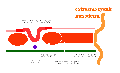
Notochord
Movie - notochord 1 | - notochord 2

The notochord is a structure which has an early mechanical role in embryonic disc folding and a major signaling role in patterning surrounding embryonic tissue development. This signaling role patterns many different tissues (neural plate, neural tube, somites, endodermal organs). It has its own sequence of development from a primitive axial process and is a developmental feature not present in the adult anatomy.
- axial process an initial epiblast hollow epithelial tube which extends in the midline from the primitive pit, cranially in the embryonic disc (toward the oral membrane).
- neuroenteric canal is a transient communication between the amnionic cavity and the yolk sac cavity formed by the axial process.
- notochordal plate forms from the axial process merging with the endoderm layer.
- notochord forms from the notochordal plate which then separates back into the mesoderm layer as a solid column of cells lying in the midline of the embryonic disc and running rostro-caudally (head to tail).
- An alternate name for the notochord is "axial mesoderm".
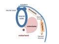
Early Placentation

The trophoblast layer has now differentiated into two morphologically distinct cellular layers.
- Syncitiotrophoblasts - form a multinucleated cytoplasmic mass by cytotrophoblast cell fusion and both invade the decidua and secrete hCG
- Cytotrophoblasts - form a cellular layer around the blastocyst, proliferates and extends behind syncitiotrophoblasts
Early Utero-Placental exchange - transfer of nutrition from maternal lacunae filled with secretions from uterine glands and maternal blood from blood vessels. The development of trophoblast villi extending into the uterine decidua.
There are three stages of villi development:
- Primary Villi - cytotrophoblast
- Secondary Villi - cytotrophoblast + extraembryonic mesoderm
- Tertiary Villi - cytotrophoblast + extraembryonic mesoderm+ blood vessels
There are two main types of early villi:
- Anchoring villi - attached to decidua
- Floating villi - not attached to decidua, floating in maternal lacunae.
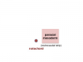
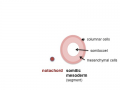
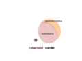
Somitogenesis


Mesoderm means the "middle layer" and it is from this layer that nearly all the bodies connective tissues are derived. In early mesoderm development a number of transient structures will form and then be lost as tissue structure is patterned and organised. Humans are vertebrates, with a "backbone", and the first mesoderm structure we will see form after the notochord will be somites.
Coelom, meaning "cavity", and major fluid-filled cavities can be seen to form both within the embryo (intraembryonic coelom) and outside the embryo (extraembryonic coelom). The intraembryonic coelom is the single primitive cavity that lies within the mesoderm layer that will eventually form the 3 major anatomical body cavities (pericardial, pleural, peritoneal).
trilaminar embryo
mesoderm regions
somite coelom
neural tube and neural crest


Somite initially forms 2 main components
- ventromedial- sclerotome forms vertebral body and intervertebral disc
- dorsolateral - dermomyotome forms dermis and skeletal muscle
paraxial mesoderm
early somite
sclerotome and dermomyotome
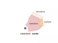
dermatome and myotome
epaxial and hypaxial muscles


![]()
Neuralation


Ectoderm - 2 parts
- midline neural plate
- columnar
- lateral surface ectoderm
- cuboidal
- sensory placodes
- epidermis of skin, hair, glands, anterior pituitary, teeth enamel
Neural Plate

- extends from buccopharyngeal membrane to primitive node
- forms above notochord and paraxial mesoderm
- neuroectodermal cells
- broad brain plate
- narrower spinal cord
- 3 components form: floor plate, neural plate, neural crest
Cardiogenesis

The Human Heart from day 10 to 25 (scanning electron micrograph)
Early Placentation

Glossary Links
- Glossary: A | B | C | D | E | F | G | H | I | J | K | L | M | N | O | P | Q | R | S | T | U | V | W | X | Y | Z | Numbers | Symbols | Term Link
- 2010 BGD: Lecture 1 | Lecture 2 | Practical 3 | Practical 6 | Practical 12
Cite this page: Hill, M.A. (2024, May 1) Embryology 2010 BGD Lecture - Development of the Embryo/Fetus 1. Retrieved from https://embryology.med.unsw.edu.au/embryology/index.php/2010_BGD_Lecture_-_Development_of_the_Embryo/Fetus_1
- © Dr Mark Hill 2024, UNSW Embryology ISBN: 978 0 7334 2609 4 - UNSW CRICOS Provider Code No. 00098G