File:Rat neural cadherin 01.jpg
Original file (1,154 × 470 pixels, file size: 62 KB, MIME type: image/jpeg)
Rat Neural Cadherin Overview
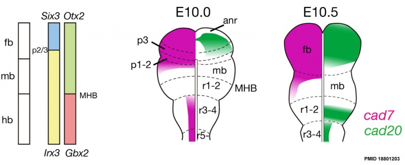
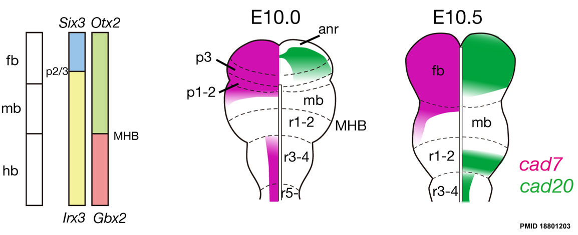
Schematic illustrations of mapping of cad7 and cad20 in the early stages.
pos, pre otic sulcus; fb, forebrain; mb, midbrain; hb, hindbrain, anr, anterior neural ridge; MHB, midbrain/hindbrain boundary.
Reference
<pubmed>18801203</pubmed> | PMC2564927 | BMC Dev Biol.
© 2008 Takahashi and Osumi; licensee BioMed Central Ltd.
This is an Open Access article distributed under the terms of the Creative Commons Attribution License (http://creativecommons.org/licenses/by/2.0), which permits unrestricted use, distribution, and reproduction in any medium, provided the original work is properly cited.
1471-213X-8-87-3.jpg
File history
Yi efo/eka'e gwa ebo wo le nyangagi wuncin ye kamina wunga tinya nan
| Gwalagizhi | Nyangagi | Dimensions | User | Comment | |
|---|---|---|---|---|---|
| current | 19:09, 23 Haziran 2012 | | 1,154 × 470 (62 KB) | Z8600021 (talk | contribs) | ==Rat Neural Cadherin Overview== Schematic illustrations of mapping of cad7 and cad20 in the early stages. pos, pre otic sulcus; fb, forebrain; mb, midbrain; hb, hindbrain, anr, anterior neural ridge; MHB, midbrain/hindbrain boundary. ===Reference=== |
You cannot overwrite this file.
File usage
The following 2 pages use this file: