File:Human week 10 fetus 04.jpg
From Embryology
Size of this preview: 800 × 600 pixels. Other resolution: 1,600 × 1,200 pixels.
Original file (1,600 × 1,200 pixels, file size: 534 KB, MIME type: image/jpeg)
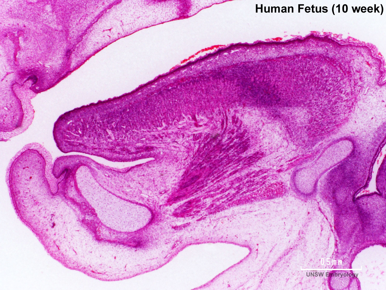
Human Female Fetus Oral Cavity (10 week)
Large image version of plane D, close to midline (H&E stain).
0.7 mm scale bar
Note: mandible development, tongue musculature
Related Images

Fetus (week 10) Planes A (most lateral), B (lateral), C (medial) and D (midline) from lateral towards the midline.
- Human Fetus - most lateral | lateral | medial | midline
- Head - most lateral | lateral | medial | midline
- Cerebellum - most lateral | lateral | medial | midline
- Urogenital Unlabelled - most lateral | lateral | medial | midline
- Urogenital Labelled - most lateral | lateral | medial | midline
- Large Images - midline
- Image Source: UNSW Embryology, no reproduction without permission.
File history
Yi efo/eka'e gwa ebo wo le nyangagi wuncin ye kamina wunga tinya nan
| Gwalagizhi | Nyangagi | Dimensions | User | Comment | |
|---|---|---|---|---|---|
| current | 17:31, 17 Haziran 2012 | | 1,600 × 1,200 (534 KB) | Z8600021 (talk | contribs) | ==Human Female Fetus Oral Cavity (10 week)== Large image version of plane D, close to midline (H&E stain). 0.7 mm scale bar Note: mandible development, tongue musculature {{10wkFetus}} |
You cannot overwrite this file.
File usage
The following 20 pages use this file:
- BGDA Practical 12 - Embryo to Fetus
- BGDB Gastrointestinal - Fetal
- Fetal Development - 10 Weeks
- Foundations Practical - Week 9 to 36
- Neural - Cranial Nerve Development
- Tongue Development
- File:Human week 10 fetus 01.jpg
- File:Human week 10 fetus 03.jpg
- File:Human week 10 fetus 04.jpg
- File:Human week 10 fetus 05.jpg
- File:Human week 10 fetus 06.jpg
- File:Human week 10 fetus 07.jpg
- File:Human week 10 fetus 08.jpg
- File:Human week 10 fetus 09.jpg
- File:Human week 10 fetus 10.jpg
- File:Human week 10 fetus 11.jpg
- File:Human week 10 fetus 12.jpg
- File:Human week 10 fetus 23.jpg
- File:Human week 10 fetus 26.jpg
- Template:Human Female Fetus Week 10 gallery


