Foundations - Histology Epithelia and Skin
Introduction


Background and Self-directed Learning for Medicine Foundations.
Practical - Histology Epithelia and Skin Virtual Slides by Patrick de Permentier.
This current page content is not part of the Foundations practical class.
Objectives
Epithelia
- Obtain an understanding of the histological appearance of various types of epithelia based on their cellular shape and number of layers.
- To examine the histological appearance of 2 other unique types of epithelia namely pseudostratified and transitional.
- To demonstrate some sites where the types of epithelia can be located
- To demonstrate certain epithelial specialisations such as microvilli and cilia.
- Relate the morphology of the epithelia to their various functions.
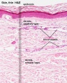
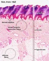
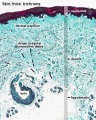
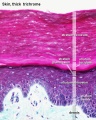
Skin
- To know the microscopic structure of the skin e.g. epidermis, dermis and hypodermis.
- To know the histological differences between hairy (thin) and glabrous (thick) skin.
- To know the histology of associated structures e.g. eccrine and apocrine sweat glands,
sebaceous glands, and hair.
- To know the histological features of some sensory receptors namely: Pacinian and Meissner
corpuscles.
Epithelia
Epithelium forms continuous layers of cells that cover surfaces and line cavities of the body.

|

|
| Epithelia cell shape | Epithelia sectioning appearance |
Epithelia Classification
- The number of cell layers: a single layer = simple epithelium; epithelia composed of more than one layer = stratified epithelia.
- The shape of the component cells when seen in sections taken at right angles to the epithelial surface: the shape may be squamous (flattened), cuboidal (about equal dimensions), or columnar (taller than it is wide).
- The presence of surface specializations e.g. cilia, microvilli and keratin.
Simple Squamous
Virtual slides: Distributing artery and vein and Aorta
Simple Cuboidal
Virtual slides: Thyroid gland and Kidney
Simple Columnar
Virtual slides: Fallopian tube-isthmus and Duodenum
Skin
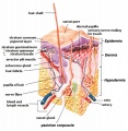
Skin structure cartoon


Terms
External Links
External Links Notice - The dynamic nature of the internet may mean that some of these listed links may no longer function. If the link no longer works search the web with the link text or name. Links to any external commercial sites are provided for information purposes only and should never be considered an endorsement. UNSW Embryology is provided as an educational resource with no clinical information or commercial affiliation.
- Blue Histology Epithelia | Skin
- UNSW Virtual Slides Medicine phase 1 (requires login for access). Histology Epithelia and Skin Virtual Slides
- UIOWA Virtual Slidebox of Histology Skin
Glossary Links
- Glossary: A | B | C | D | E | F | G | H | I | J | K | L | M | N | O | P | Q | R | S | T | U | V | W | X | Y | Z | Numbers | Symbols | Term Link
Cite this page: Hill, M.A. (2024, June 17) Embryology Foundations - Histology Epithelia and Skin. Retrieved from https://embryology.med.unsw.edu.au/embryology/index.php/Foundations_-_Histology_Epithelia_and_Skin
- © Dr Mark Hill 2024, UNSW Embryology ISBN: 978 0 7334 2609 4 - UNSW CRICOS Provider Code No. 00098G