Category:Carnegie Stage 7
From Embryology
Subcategories
This category has the following 9 subcategories, out of 9 total.
Pages in category 'Carnegie Stage 7'
The following 55 pages are in this category, out of 55 total.
C
P
- Paper - A human embryo of the pre-somite period from the uterine tube
- Paper - A human embryo with head-process and commencing arch enteric canal
- Paper - A Note on the Development of the Septum Transversum and the Liver
- Paper - A presomite human embryo showing a yolk-sac duct
- Paper - A presomite human embryo showing an early stage of the primitive streak
- Paper - An early human embryo (no. 1285, Manchester Collection) with capsular attachment of the connecting stalk (1935)
- Paper - An Early Human Embryo (No. 1285, Manchester Collection), with Capsular Attachment of the Connecting Stalk
- Paper - An Early Human Embryo, with 0.55 mm long Embryonic Shield
- Paper - Description of a Human Embryo of Twenty-three Paired Somites
- Paper - Normal development of early human embryos: Observation of 90 specimens at Carnegie stages 7 to 13
- Paper - Six normal and complete presomite human ova
- Paper - The Falkiner ovum
- Paper - Two Early Human Embryos
- Paper - Two presomite human embryos
R
- Template:Ref-Brewer1937
- Template:Ref-BrewerFitzgerald1937
- Template:Ref-FlorianHill1935
- Template:Ref-George1942
- Template:Ref-Kindred1933
- Template:Ref-Mazanec1949
- Template:Ref-Mazanec1959
- Template:Ref-PMID13044724
- Template:Ref-Stieve1926a
- Template:Ref-Stieve1926b
- Template:Ref-Thompson1923
- Template:Ref-West1952
S
Media in category 'Carnegie Stage 7'
The following 84 files are in this category, out of 84 total.
 Florian1935 chorionic vesicle fig11.jpg 1,009 × 952; 248 KB
 Florian1935 fig01.jpg 774 × 405; 115 KB
 Florian1935 fig02.jpg 790 × 405; 117 KB
 Florian1935 fig03.jpg 800 × 455; 130 KB
 Florian1935 fig04.jpg 798 × 443; 120 KB
 Florian1935 fig05.jpg 803 × 227; 58 KB
 Florian1935 fig06.jpg 770 × 573; 145 KB
 Florian1935 fig07.jpg 597 × 812; 206 KB
 Florian1935 fig08.jpg 691 × 480; 114 KB
 Florian1935 fig09.jpg 510 × 765; 168 KB
 Florian1935 fig10.jpg 464 × 721; 157 KB
 Florian1935 fig11.jpg 1,280 × 1,427; 443 KB
 Florian1935 plate01.jpg 1,481 × 1,825; 529 KB
 Florian1935 plate02.jpg 1,481 × 1,806; 578 KB
 Florian1935 plate03.jpg 1,481 × 2,000; 735 KB
 Florian1935 textfig01.jpg 1,120 × 1,202; 136 KB
 Florian1935 textfig02.jpg 1,579 × 2,000; 342 KB
 Florian1935 textfig03.jpg 1,579 × 2,000; 212 KB
 MartinFalkiner1938 fig01.jpg 800 × 931; 133 KB
 MartinFalkiner1938 fig02.jpg 600 × 886; 76 KB
 MartinFalkiner1938 fig03.jpg 700 × 1,000; 89 KB
 MartinFalkiner1938 fig04.jpg 700 × 974; 82 KB
 MartinFalkiner1938 fig05.jpg 700 × 1,022; 88 KB
 MartinFalkiner1938 fig06.jpg 700 × 1,009; 87 KB
 MartinFalkiner1938 fig07.jpg 700 × 951; 63 KB
 MartinFalkiner1938 fig08.jpg 670 × 840; 46 KB
 MartinFalkiner1938 plate01.jpg 1,280 × 1,803; 344 KB
 MartinFalkiner1938 plate02.jpg 1,280 × 1,840; 247 KB
 Morton1949 fig01.jpg 1,673 × 1,282; 615 KB
 Morton1949 fig02.jpg 652 × 786; 79 KB
 Morton1949 fig03.jpg 1,016 × 786; 108 KB
 Morton1949 fig04.jpg 629 × 1,316; 332 KB
 Morton1949 fig05.jpg 738 × 1,316; 383 KB
 Morton1949 fig06.jpg 1,012 × 868; 253 KB
 Morton1949 plate01.jpg 1,784 × 2,457; 975 KB
 Morton1949 text-fig01.jpg 847 × 1,084; 111 KB
 Morton1949 text-fig02.jpg 778 × 1,078; 70 KB
 Stage7 800x700px.jpg 690 × 800; 69 KB
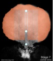
 Stage7 axes.jpg 500 × 375; 9 KB
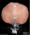
 Stage7 axial process.jpg 500 × 375; 11 KB
 Stage7 bf5.jpg 1,712 × 1,206; 875 KB
 Stage7 bf51.jpg 600 × 450; 96 KB
 Stage7 bf52.jpg 600 × 450; 114 KB
 Stage7 bf53.jpg 500 × 375; 74 KB
 Stage7 bf5a.jpg 1,024 × 721; 690 KB
 Stage7 bf5b.jpg 500 × 352; 216 KB
 Stage7 bf6.jpg 347 × 599; 69 KB
 Stage7 bf7.jpg 400 × 341; 22 KB
 Stage7 bf8.jpg 400 × 341; 18 KB
 Stage7 bf9.jpg 1,200 × 1,039; 501 KB
 Stage7 cloacal-oral-membranes.jpg 690 × 800; 70 KB
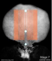
 Stage7 features.jpg 500 × 375; 9 KB
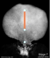
 Stage7 folding.jpg 500 × 375; 10 KB
 Stage7 intermediate-mesoderm.jpg 690 × 800; 67 KB
 Stage7 lateral-plate.jpg 690 × 800; 65 KB
 Stage7 mesoderm.jpg 690 × 800; 67 KB
 Stage7 notochord.jpg 690 × 800; 70 KB
 Stage7 paraxial-mesoderm.jpg 690 × 800; 69 KB
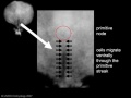 Stage7 primitive streak labelled.jpg 500 × 375; 13 KB
 Stage7 primitive-streak-node.jpg 690 × 800; 69 KB
 Stage7-bf1.jpg 600 × 676; 34 KB
 Stage7-bf2.jpg 800 × 579; 53 KB
 Stage7-bf3.jpg 523 × 600; 45 KB
 Stage7-bf4.jpg 800 × 695; 53 KB
 Stage7-sem1.jpg 814 × 600; 100 KB
 Stage7-sem2.jpg 590 × 800; 98 KB
 Stage7-sem4.jpg 937 × 595; 79 KB
 Stage7-sem5.jpg 1,000 × 735; 202 KB
 Stage7.jpg 312 × 427; 7 KB
 ThompsonBrash1923 fig01.jpg 961 × 917; 212 KB
 ThompsonBrash1923 fig02.jpg 910 × 1,345; 146 KB
 ThompsonBrash1923 fig03.jpg 1,327 × 1,131; 135 KB
 ThompsonBrash1923 fig04.jpg 1,453 × 806; 166 KB
 ThompsonBrash1923 fig05.jpg 1,420 × 655; 226 KB
 ThompsonBrash1923 fig06.jpg 1,348 × 865; 166 KB
 ThompsonBrash1923 fig07.jpg 1,440 × 872; 324 KB
 ThompsonBrash1923 fig08.jpg 1,395 × 1,222; 299 KB
 ThompsonBrash1923 fig09.jpg 1,145 × 1,151; 518 KB
 ThompsonBrash1923 fig10.jpg 1,342 × 875; 249 KB
 ThompsonBrash1923 fig11.jpg 1,336 × 1,344; 385 KB
 ThompsonBrash1923 fig12.jpg 1,018 × 680; 160 KB
 West1952 plate01.jpg 1,280 × 1,797; 251 KB
 West1952 plate02.jpg 1,280 × 1,651; 284 KB
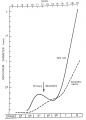 Yolk sac and amniotic cavity volume graph.jpg 719 × 1,000; 50 KB


