File:Ovary histology 003.jpg

Original file (1,280 × 1,024 pixels, file size: 337 KB, MIME type: image/jpeg)
Ovary - Corpus albicans
Hormone secretion in the corpus luteum (CL) ceases within 14 days after ovulation if fertilisation and implantation have not occurred.
- With no implantation and development, the corpus luteum degenerates into a corpus albicans, a whitish scar tissue within the ovary.
- With implantation and development, syncytiotrophoblast commence secretion of human chorionic gonadotropin (hCG) that hormonally supports the CL and the corpus albicans does not form.
Hormone secretion continues for 2-3 month after ovulation if fertilisation occurs.
Ovary, monkey H&E reproductive system, female, corpus albicans, primary follicle, primordial follicle, granulosa cells, oocyte x20
Ovary histology: Tunica Albuginea x20 | Tunica albuginea, Germinal epithelium x40 | Primary follicle, primordial follicle, oocyte, x40 | Secondary follicle, cumulus oophorus, zona pelucida, granulosa cells, oocyte x20 | Corpus luteum, theca lutein cells, granulosa lutein cells, Loupe | Corpus luteum, theca lutein cells, granulosa lutein cells, x10 | Corpus luteum, theca lutein cells, granulosa lutein cells, x40 | Corpus albicans, primary follicle, primordial follicle, granulosa cells, oocyte x20 | Menstrual Cycle | Ovary Development
Links: Histology | Histology Stains | Blue Histology images copyright Lutz Slomianka 1998-2009. The literary and artistic works on the original Blue Histology website may be reproduced, adapted, published and distributed for non-commercial purposes. See also the page Histology Stains.
Cite this page: Hill, M.A. (2024, June 21) Embryology Ovary histology 003.jpg. Retrieved from https://embryology.med.unsw.edu.au/embryology/index.php/File:Ovary_histology_003.jpg
- © Dr Mark Hill 2024, UNSW Embryology ISBN: 978 0 7334 2609 4 - UNSW CRICOS Provider Code No. 00098G
File:Ovary_histology_003.jpg
File history
Yi efo/eka'e gwa ebo wo le nyangagi wuncin ye kamina wunga tinya nan
| Gwalagizhi | Nyangagi | Dimensions | User | Comment | |
|---|---|---|---|---|---|
| current | 21:08, 23 February 2011 | | 1,280 × 1,024 (337 KB) | S8600021 (talk | contribs) | File:Ovary_histology_003.jpg Ovary,_monkey_H&E_reproductive_system,_female,_corpus_albicans,_primary_follicle,_primordial_follicle,_granulosa_cells,_oocyte_x20.jpg {{Ovary Histology}} {{Template:Blue Histology}} Category:Monkey [[Category:Genital |
| 21:08, 23 February 2011 | 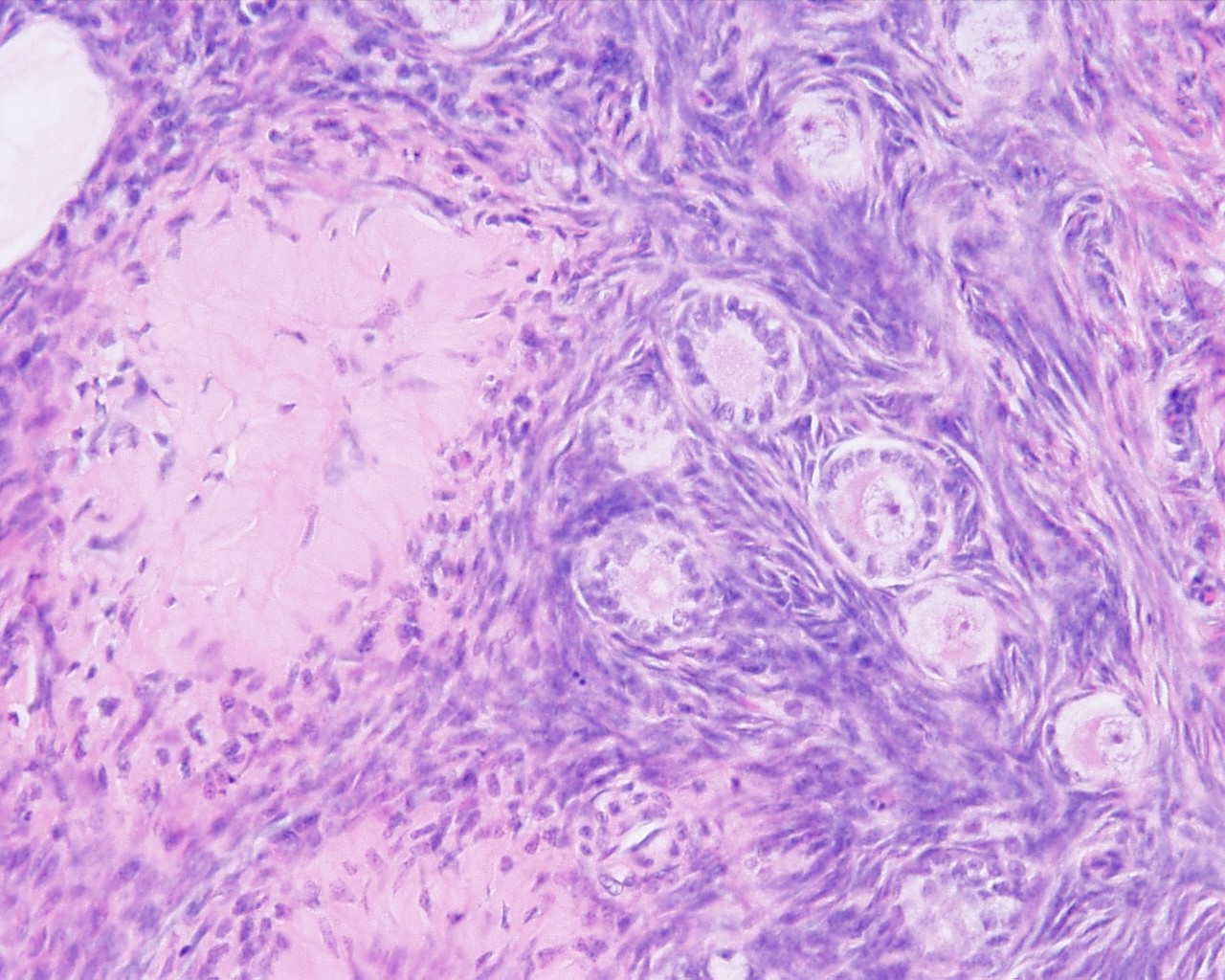 | 1,280 × 1,024 (337 KB) | S8600021 (talk | contribs) | File:Ovary_histology_003.jpg Ovary,_monkey_H&E_reproductive_system,_female,_corpus_albicans,_primary_follicle,_primordial_follicle,_granulosa_cells,_oocyte_x20.jpg {{Ovary Histology}} {{Template:Blue Histology}} Category:Monkey [[Category:Genita |
You cannot overwrite this file.