File:Skull CT normal sutures.jpg
Original file (1,000 × 900 pixels, file size: 138 KB, MIME type: image/jpeg)
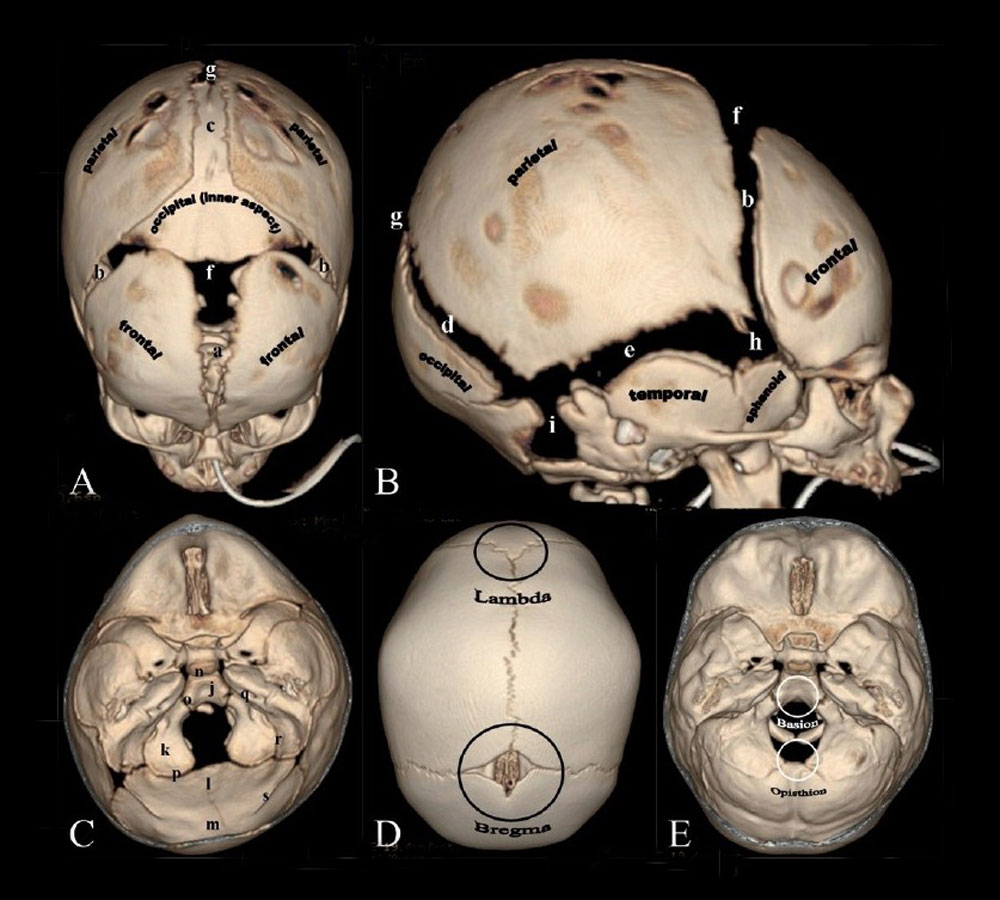
Skull Normal Sutures
Computed Tomography (CT) scan with 3D surface-rendered reconstructions.
Vertex (A) and lateral (B) views.
- (a) Metopic suture; (b) coronal sutures; (c) sagittal suture; (d) lambdoid suture; (e) squamosal suture; (f) anterior fontanel; (g) posterior fontanel; (h) sphenoidal fontanel; (i) mastoid fontanel.
- Cranial vault bones usually ossify from the center to periphery, which results in this “widened” appearance of the sutures in the newborn.
Endocranial skull base view (C) shows portions of the occipital bone and sutures
- (j) Basioccipital; (k) paired exoccipital; (l) supraoccipital; and (m) interparietal. Associated synchondroses are (n) spheno-occipital; (o)anterior intra-occipital; (p) posterior intra-occipital; (q) petro-occipital; (r) occipitomastoid; (s) and mendosal sutures. Note that o, k, p and s are paired structures.
Vertex view (D) shows the lambda (point of intersection of the sagittal and lambdoid sutures) and bregma (point of intersection of the coronal and sagittal sutures.
Endocranial skull base view (E) shows the basion (located on the basiocciput, at the midpoint of the anterior margin of the foramen magnum) and opisthion (located on the occipital bone, at the midpoint of the posterior margin of the foramen magnum).
Original file name: Figure 1(A-E): IJRI-21-49-g001.jpg
Reference
<pubmed>21431034</pubmed>| PMC3056371 | Indian J Radiol Imaging.
This is an open-access article distributed under the terms of the Creative Commons Attribution License, which permits unrestricted use, distribution, and reproduction in any medium, provided the original work is properly cited.
File history
Yi efo/eka'e gwa ebo wo le nyangagi wuncin ye kamina wunga tinya nan
| Gwalagizhi | Nyangagi | Dimensions | User | Comment | |
|---|---|---|---|---|---|
| current | 08:01, 17 March 2012 | | 1,000 × 900 (138 KB) | Z8600021 (talk | contribs) | |
| 18:33, 23 May 2011 |  | 750 × 689 (105 KB) | S8600021 (talk | contribs) | ==Skull Normal Sutures== Computed Tomography (CT) scan with 3D surface-rendered reconstructions. Vertex (A) and lateral (B) views. (a) Metopic suture; (b) coronal sutures; (c) sagittal suture; (d) lambdoid suture; (e) squamosal suture; (f) anterior fonta |
You cannot overwrite this file.
File usage
The following 10 pages use this file:
- 2011 Lab 6 - Postnatal
- AACP Meeting 2013 - Face Embryology
- ANAT2341 Lab 6 - Postnatal
- BGDB Face and Ear - Postnatal
- BGD Lecture - Face and Ear Development
- Computed Tomography
- Head Development
- Lecture - Head Development
- Musculoskeletal System - Skull Development
- Neural Exam - Newborn head shape and sutures