File:Bladder histology.jpg
Bladder_histology.jpg (300 × 400 pixels, file size: 56 KB, MIME type: image/jpeg)
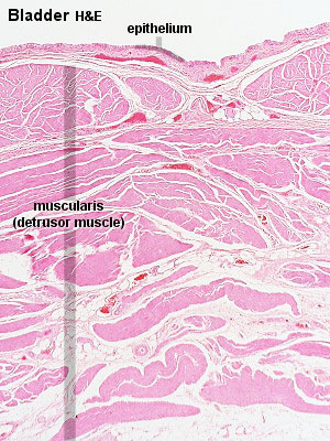
Bladder Histology
- transitional epithelium
- epithelium is smooth, without appreciable crypts or folds.
Although the precise orientation of the muscles in the wall of the bladder depends on where the tissue block was taken and at which angle it was sectioned, it should be possible to identify three tiers of muscle bundles at least in some parts of the bladder wall.
Original File name: Bla02he-1.jpg Source: UWA Blue Histology
Links: Histology | Histology Stains | Blue Histology images copyright Lutz Slomianka 1998-2009. The literary and artistic works on the original Blue Histology website may be reproduced, adapted, published and distributed for non-commercial purposes. See also the page Histology Stains.
Cite this page: Hill, M.A. (2026, October 2) Embryology Bladder histology.jpg. Retrieved from https://embryology.med.unsw.edu.au/embryology/index.php/File:Bladder_histology.jpg
- © Dr Mark Hill 2026, UNSW Embryology ISBN: 978 0 7334 2609 4 - UNSW CRICOS Provider Code No. 00098G
File history
Yi efo/eka'e gwa ebo wo le nyangagi wuncin ye kamina wunga tinya nan
| Gwalagizhi | Nyangagi | Dimensions | User | Comment | |
|---|---|---|---|---|---|
| current | 14:10, 19 September 2009 | | 300 × 400 (56 KB) | S8600021 (talk | contribs) | bladder histology Original File name: Bla02he-1.jpg Source: UWA Blue Histology |
You cannot overwrite this file.
File usage
The following 7 pages use this file:


