File:Congdon1922-plate03.jpg
From Embryology
Size of this preview: 800 × 590 pixels. Other resolution: 1,200 × 885 pixels.
Original file (1,200 × 885 pixels, file size: 182 KB, MIME type: image/jpeg)
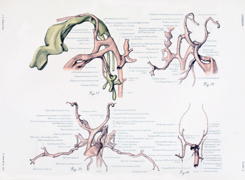
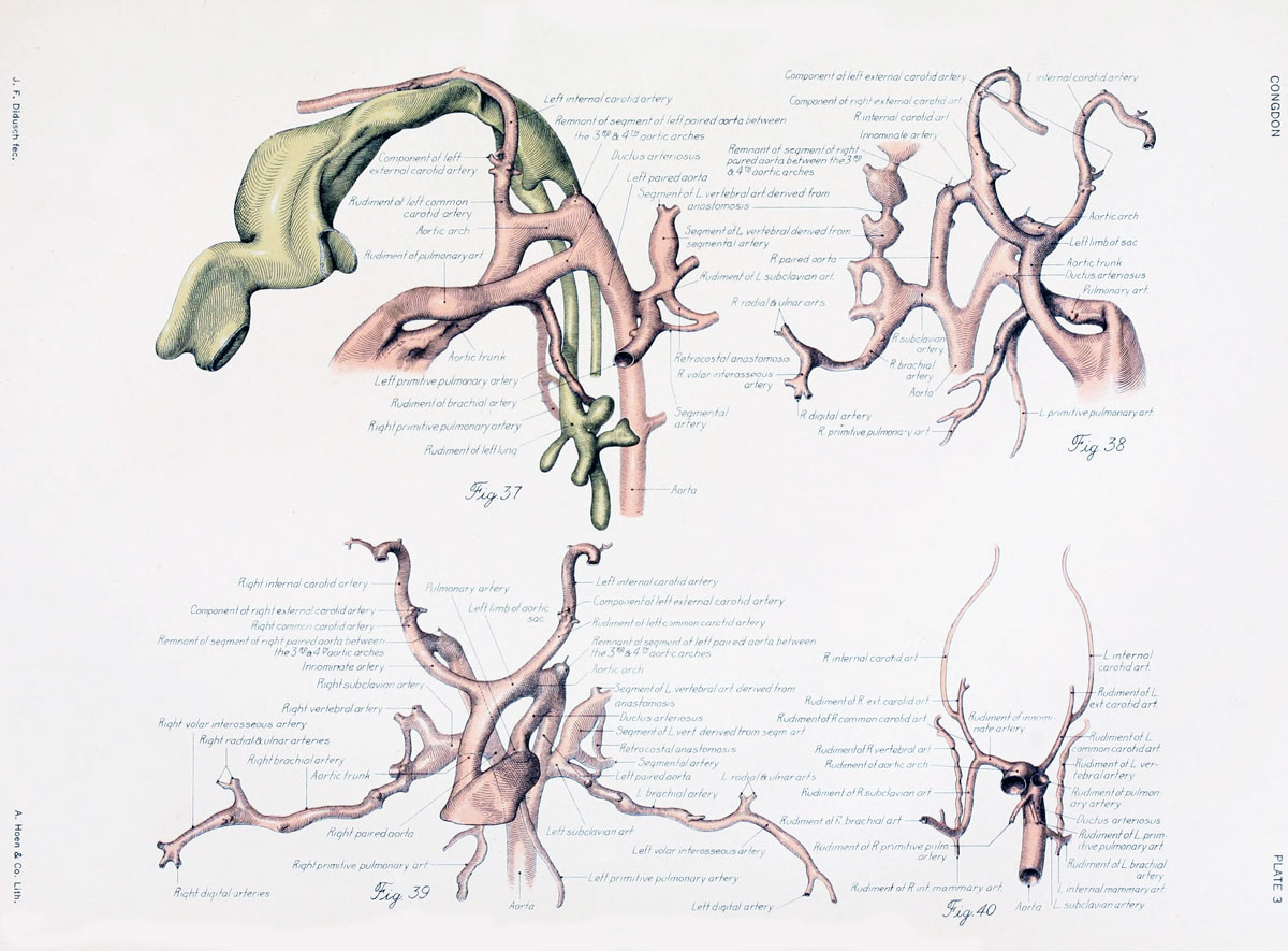
Plate 3
Figs. 37 to 39. A 14-mm. embryo
(No. 940) in which the last indications of the aortic-arch system are just disappearing and a very primitive condition of the larger arteries derived from them is already recognizable. Figure 37, lateral view; figure 38 ventrolateral view; figure 39, ventral view.
Fig. 40. Ventral view of an 18-mm. embryo
(No. 1390). The arterial evolution has preceded so far that the adult vessels are easily recognizable.
| Historic Disclaimer - information about historic embryology pages |
|---|
 |
File history
Yi efo/eka'e gwa ebo wo le nyangagi wuncin ye kamina wunga tinya nan
| Gwalagizhi | Nyangagi | Dimensions | User | Comment | |
|---|---|---|---|---|---|
| current | 18:01, 1 September 2012 | | 1,200 × 885 (182 KB) | Z8600021 (talk | contribs) | |
| 19:20, 7 Mayıs 2011 |  | 1,173 × 1,011 (169 KB) | S8600021 (talk | contribs) | ==Plate 3== ===Figs. 37 to 39. A 14-mm. embryo=== (No. 940) in which the last indications of the aortic-arch system are just disappearing and a very primitive condition of the larger arteries derived from them is already recognizable. Figure 37, lateral |
You cannot overwrite this file.