File:Congdon1922-plate01.jpg
From Embryology
Size of this preview: 438 × 599 pixels. Other resolution: 877 × 1,200 pixels.
Original file (877 × 1,200 pixels, file size: 145 KB, MIME type: image/jpeg)
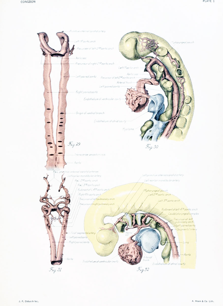
Plate 1
Figs. 29 and 30
Ventral and lateral views of the cranial portion of the arterial system of a 22-somite embryo. The first arch is at its maximum development and the dorsal and ventral outgrowths, which are to aid in the formation of the second, are just appearing. Embryo No. 2053, length 3 mm.
Figs. 31 and 32
Ventral and lateral views of an embryo in which the first arch has gone, the second arch is much reduced in diameter, and the third arch well developed. Dorsal and ventral outgrowths for the fourth and probably for the pulmonary arch are present. Embryo No. 836, length 4 mm.
| Historic Disclaimer - information about historic embryology pages |
|---|
 |
File history
Yi efo/eka'e gwa ebo wo le nyangagi wuncin ye kamina wunga tinya nan
| Gwalagizhi | Nyangagi | Dimensions | User | Comment | |
|---|---|---|---|---|---|
| current | 16:38, 1 September 2012 | | 877 × 1,200 (145 KB) | Z8600021 (talk | contribs) | |
| 19:09, 7 Mayıs 2011 | 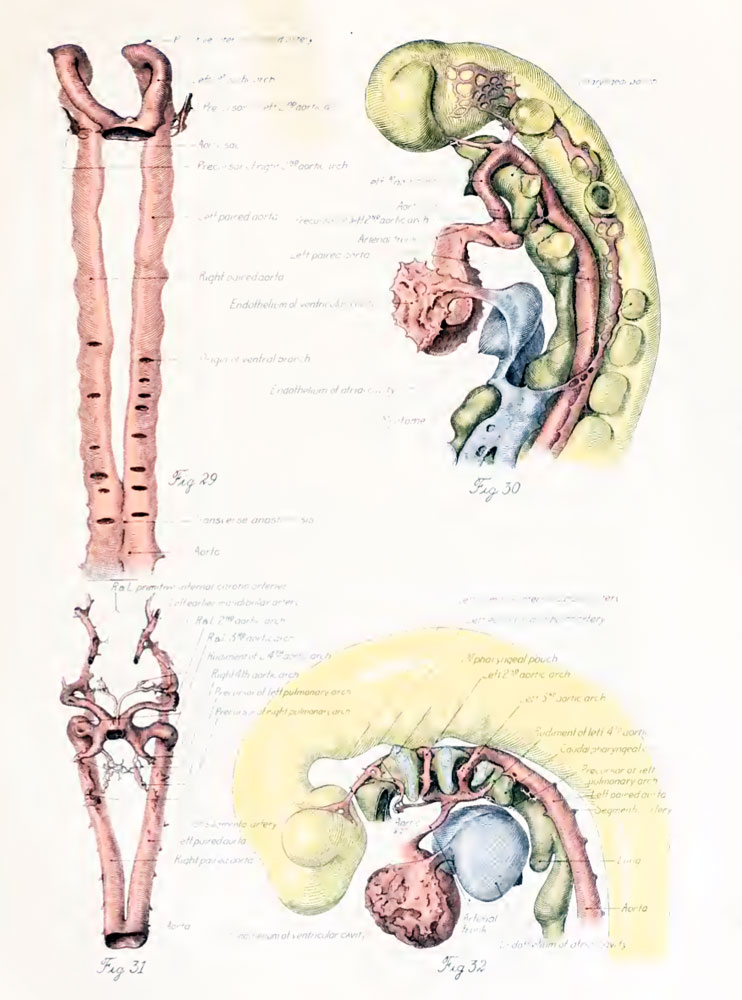 | 742 × 1,000 (116 KB) | S8600021 (talk | contribs) | ==Plate 1== ===Figs. 29 and 30=== Ventral and lateral views of the cranial portion of the arterial system of a 22-somite embryo. The first arch is at its maximum development and the dorsal and ventral outgrowths, which are to aid in the formation of the |
You cannot overwrite this file.


